|
|
|
|
 Description
|
|
Compounds
|
||||||||||||||||||||||||||||||||||||||||||||||||
Chains, Units
Summary Information (see also Sequences/Alignments below) |
Ligands, Modified Residues, Ions (2, 8)| Asymmetric/Biological Unit (2, 8) |
Sites (2, 2)
Asymmetric Unit (2, 2)
|
SS Bonds (0, 0)| (no "SS Bond" information available for 2V8L) |
Cis Peptide Bonds (0, 0)| (no "Cis Peptide Bond" information available for 2V8L) |
SAPs(SNPs)/Variants (0, 0)| (no "SAP(SNP)/Variant" information available for 2V8L) |
PROSITE Motifs (0, 0)| (no "PROSITE Motif" information available for 2V8L) |
Exons (0, 0)| (no "Exon" information available for 2V8L) |
Sequences/Alignments
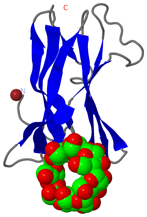
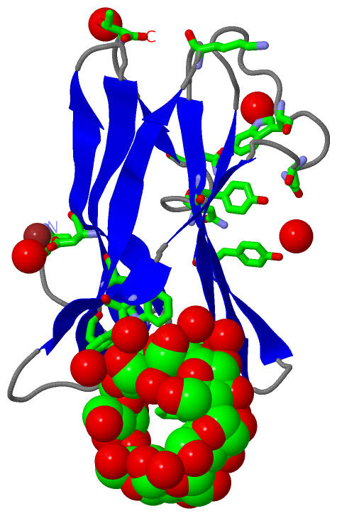
Asymmetric/Biological UnitChain A from PDB Type:PROTEIN Length:106 aligned with B7XC04_RHIOR | B7XC04 from UniProtKB/TrEMBL Length:604 Alignment length:106 35 45 55 65 75 85 95 105 115 125 B7XC04_RHIOR 26 ASIPSSASVQLDSYNYDGSTFSGKIYVKNIAYSKKVTVVYADGSDNWNNNGNIIAASFSGPISGSNYEYWTFSASVKGIKEFYIKYEVSGKTYYDNNNSANYQVST 131 SCOP domains ---------------------------------------------------------------------------------------------------------- SCOP domains CATH domains ---------------------------------------------------------------------------------------------------------- CATH domains Pfam domains ---------------------------------------------------------------------------------------------------------- Pfam domains SAPs(SNPs) ---------------------------------------------------------------------------------------------------------- SAPs(SNPs) PROSITE ---------------------------------------------------------------------------------------------------------- PROSITE Transcript ---------------------------------------------------------------------------------------------------------- Transcript 2v8l A 1 ASIPSSASVQLDSYNYDGSTFSGKIYVKNIAYSKKVTVVYADGSDNWNNNGNIIAASFSGPISGSNYEYWTFSASVKGIKEFYIKYEVSGKTYYDNNNSANYQVST 106 10 20 30 40 50 60 70 80 90 100 Chain A from PDB Type:PROTEIN Length:106 aligned with Q2VC81_RHIOR | Q2VC81 from UniProtKB/TrEMBL Length:604 Alignment length:106 35 45 55 65 75 85 95 105 115 125 Q2VC81_RHIOR 26 ASIPSSASVQLDSYNYDGSTFSGKIYVKNIAYSKKVTVVYADGSDNWNNNGNTIAASFSGPISGSNYEYWTFSASVKGIKEFYIKYEVSGKTYYDNNNSANYQVST 131 SCOP domains ---------------------------------------------------------------------------------------------------------- SCOP domains CATH domains ---------------------------------------------------------------------------------------------------------- CATH domains Pfam domains ---------------------------------------------------------------------------------------------------------- Pfam domains SAPs(SNPs) ---------------------------------------------------------------------------------------------------------- SAPs(SNPs) PROSITE ---------------------------------------------------------------------------------------------------------- PROSITE Transcript ---------------------------------------------------------------------------------------------------------- Transcript 2v8l A 1 ASIPSSASVQLDSYNYDGSTFSGKIYVKNIAYSKKVTVVYADGSDNWNNNGNIIAASFSGPISGSNYEYWTFSASVKGIKEFYIKYEVSGKTYYDNNNSANYQVST 106 10 20 30 40 50 60 70 80 90 100
|
||||||||||||||||||||
SCOP Domains (0, 0)| (no "SCOP Domain" information available for 2V8L) |
CATH Domains (0, 0)| (no "CATH Domain" information available for 2V8L) |
Pfam Domains (0, 0)| (no "Pfam Domain" information available for 2V8L) |
Gene Ontology (4, 7)|
Asymmetric/Biological Unit(hide GO term definitions) Chain A (Q2VC81_RHIOR | Q2VC81)
Chain A (B7XC04_RHIOR | B7XC04)
|
||||||||||||||||||||||||||||||||||||||||||||||||||||||||||||||||||
Interactive Views
|
||||||||||||||||||||||||||||||||||||||||||||||||||||||||||||||||||||||||||||||||||||||||||||||||||||||||||||||||||||||||||||||||||||
Still Images
|
||||||||||||||||
Databases
|
||||||||||||||||||||||||||||||||||||||||||||||||||||||||||||||||||||||||||||||||||||||||||||||||||||||||||||||||||||||||||||||||||||||||||||||||||||||||||||||||||||||||||||||||||||||||||
Analysis Tools
|
||||||||||||||||||||||||||||||||||||||||||||||||||||||||||||||||||||||||
Entries Sharing at Least One Protein Chain (UniProt ID)
Related Entries Specified in the PDB File
|
|