Show PDB file:   
         Plain Text   HTML   (compressed file size)
QuickSearch:   
by PDB,NDB,UniProt,PROSITE Code or Search Term(s)  
(-)NMR Structure - manually
(-)NMR Structure - model 1
(-)NMR Structure - all models
collapse expand < >
Image NMR Structure - manually
NMR Structure - manually  (Jmol Viewer)
Image NMR Structure - model 1
NMR Structure - model 1  (Jmol Viewer)
Image NMR Structure - all models
NMR Structure - all models  (Jmol Viewer)

(-) Description

Title :  SOLUTION STRUCTURE OF ASPP2 N-TERMINUS
 
Authors :  H. Tidow, T. J. Rutherford, A. Andreeva, A. R. Fersht
Date :  22 Mar 07  (Deposition) - 10 Jul 07  (Release) - 24 Feb 09  (Revision)
Method :  SOLUTION NMR
Resolution :  NOT APPLICABLE
Chains :  NMR Structure  :  A  (20x)
Keywords :  Aspp2, Ubiquitin-Like, Sh3-Domain, Apoptosis, Cell Cycle, Ank Repeat, Sh3-Binding (Keyword Search: [Gene Ontology, PubMed, Web (Google))
 
Reference :  H. Tidow, A. Andreeva, T. J. Rutherford, A. R. Fersht
Solution Structure Of Aspp2 N-Terminal Domain (N- Aspp2) Reveals A Ubiquitin-Like Fold.
J. Mol. Biol. V. 371 948 2007
PubMed-ID: 17594908  |  Reference-DOI: 10.1016/J.JMB.2007.05.024

(-) Compounds

Molecule 1 - APOPTOSIS-STIMULATING OF P53 PROTEIN 2
    ChainsA
    EngineeredYES
    Expression SystemESCHERICHIA COLI
    Expression System StrainBL21
    Expression System Taxid511693
    Expression System VectorPRSET
    FragmentRESIDUES 1-83
    Organism CommonHUMAN
    Organism ScientificHOMO SAPIENS
    Organism Taxid9606
    SynonymTUMOR SUPPRESSOR P53-BINDING PROTEIN 2,P53-BINDING PROTEIN 2,P53BP2,53BP2, BCL2-BINDING PROTEIN, BBP, RENAL CARCINOMA ANTIGEN NY-REN-51

 Structural Features

(-) Chains, Units

  
NMR Structure (20x)

Summary Information (see also Sequences/Alignments below)

(-) Ligands, Modified Residues, Ions  (0, 0)

(no "Ligand,Modified Residues,Ions" information available for 2UWQ)

(-) Sites  (0, 0)

(no "Site" information available for 2UWQ)

(-) SS Bonds  (0, 0)

(no "SS Bond" information available for 2UWQ)

(-) Cis Peptide Bonds  (0, 0)

(no "Cis Peptide Bond" information available for 2UWQ)

 Sequence-Structure Mapping

(-) SAPs(SNPs)/Variants  (0, 0)

(no "SAP(SNP)/Variant" information available for 2UWQ)

(-) PROSITE Motifs  (0, 0)

(no "PROSITE Motif" information available for 2UWQ)

(-) Exons   (0, 0)

(no "Exon" information available for 2UWQ)

(-) Sequences/Alignments

NMR Structure
   Reformat: Number of residues per line =  ('0' or empty: single-line sequence representation)
  Number of residues per labelling interval =   
  UniProt sequence: complete  aligned part    
   Show mapping: SCOP domains CATH domains Pfam domains Secondary structure (by author)
SAPs(SNPs) PROSITE motifs Exons
(details for a mapped element are shown in a popup box when the mouse pointer rests over it)
Chain A from PDB  Type:PROTEIN  Length:86
 aligned with ASPP2_HUMAN | Q13625 from UniProtKB/Swiss-Prot  Length:1128

    Alignment length:86
                               1                                                                                  
                               |     7        17        27        37        47        57        67        77      
           ASPP2_HUMAN    - ---MMPMFLTVYLSNNEQHFTEVPVTPETICRDVVDLCKEPGESDCHLAEVWCGSERPVADNERMFDVLQRFGSQRNEVRFFLRHE 83
               SCOP domains -------------------------------------------------------------------------------------- SCOP domains
               CATH domains -------------------------------------------------------------------------------------- CATH domains
               Pfam domains -------------------------------------------------------------------------------------- Pfam domains
         Sec.struct. author ......eeeeee.......eeeeee.....hhhhhhhh........eeeeee..eeeee....hhhhhhhhhh.hhhhheeeee.. Sec.struct. author
                 SAPs(SNPs) -------------------------------------------------------------------------------------- SAPs(SNPs)
                    PROSITE -------------------------------------------------------------------------------------- PROSITE
                 Transcript -------------------------------------------------------------------------------------- Transcript
                  2uwq A -2 GGSMMPMFLTVYLSNNEQHFTEVPVTPETICRDVVDLCKEPGESDCHLAEVWCGSERPVADNERMFDVLQRFGSQRNEVRFFLRHE 83
                                     7        17        27        37        47        57        67        77      

   Legend:   → Mismatch (orange background)
  - → Gap (green background, '-', border residues have a numbering label)
    → Modified Residue (blue background, lower-case, 'x' indicates undefined single-letter code, labelled with number + name)
  x → Chemical Group (purple background, 'x', labelled with number + name, e.g. ACE or NH2)
  extra numbering lines below/above indicate numbering irregularities and modified residue names etc., number ends below/above '|'

 Classification and Annotation

(-) SCOP Domains  (0, 0)

(no "SCOP Domain" information available for 2UWQ)

(-) CATH Domains  (0, 0)

(no "CATH Domain" information available for 2UWQ)

(-) Pfam Domains  (0, 0)

(no "Pfam Domain" information available for 2UWQ)

(-) Gene Ontology  (21, 21)

NMR Structure(hide GO term definitions)
Chain A   (ASPP2_HUMAN | Q13625)
molecular function
    GO:0051059    NF-kappaB binding    Interacting selectively and non-covalently with NF-kappaB, a transcription factor for eukaryotic RNA polymerase II promoters.
    GO:0017124    SH3 domain binding    Interacting selectively and non-covalently with a SH3 domain (Src homology 3) of a protein, small protein modules containing approximately 50 amino acid residues found in a great variety of intracellular or membrane-associated proteins.
    GO:0005070    SH3/SH2 adaptor activity    Interacting selectively and non-covalently and simultaneously with one or more signal transduction molecules, usually acting as a scaffold to bring these molecules into close proximity either using their own SH2/SH3 domains (e.g. Grb2) or those of their target molecules (e.g. SAM68).
    GO:0042802    identical protein binding    Interacting selectively and non-covalently with an identical protein or proteins.
    GO:0002039    p53 binding    Interacting selectively and non-covalently with one of the p53 family of proteins.
    GO:0005515    protein binding    Interacting selectively and non-covalently with any protein or protein complex (a complex of two or more proteins that may include other nonprotein molecules).
biological process
    GO:0006915    apoptotic process    A programmed cell death process which begins when a cell receives an internal (e.g. DNA damage) or external signal (e.g. an extracellular death ligand), and proceeds through a series of biochemical events (signaling pathway phase) which trigger an execution phase. The execution phase is the last step of an apoptotic process, and is typically characterized by rounding-up of the cell, retraction of pseudopodes, reduction of cellular volume (pyknosis), chromatin condensation, nuclear fragmentation (karyorrhexis), plasma membrane blebbing and fragmentation of the cell into apoptotic bodies. When the execution phase is completed, the cell has died.
    GO:0007049    cell cycle    The progression of biochemical and morphological phases and events that occur in a cell during successive cell replication or nuclear replication events. Canonically, the cell cycle comprises the replication and segregation of genetic material followed by the division of the cell, but in endocycles or syncytial cells nuclear replication or nuclear division may not be followed by cell division.
    GO:0072332    intrinsic apoptotic signaling pathway by p53 class mediator    A series of molecular signals in which an intracellular signal is conveyed to trigger the apoptotic death of a cell. The pathway is induced by the cell cycle regulator phosphoprotein p53, or an equivalent protein, and ends when the execution phase of apoptosis is triggered.
    GO:0045786    negative regulation of cell cycle    Any process that stops, prevents or reduces the rate or extent of progression through the cell cycle.
    GO:1900119    positive regulation of execution phase of apoptosis    Any process that activates or increases the frequency, rate or extent of execution phase of apoptosis.
    GO:1901216    positive regulation of neuron death    Any process that activates or increases the frequency, rate or extent of neuron death.
    GO:1900740    positive regulation of protein insertion into mitochondrial membrane involved in apoptotic signaling pathway    Any process that activates or increases the frequency, rate or extent of protein insertion into mitochondrial membrane involved in apoptotic signaling pathway.
    GO:0042981    regulation of apoptotic process    Any process that modulates the occurrence or rate of cell death by apoptotic process.
    GO:1901796    regulation of signal transduction by p53 class mediator    Any process that modulates the frequency, rate or extent of signal transduction by p53 class mediator.
    GO:0007165    signal transduction    The cellular process in which a signal is conveyed to trigger a change in the activity or state of a cell. Signal transduction begins with reception of a signal (e.g. a ligand binding to a receptor or receptor activation by a stimulus such as light), or for signal transduction in the absence of ligand, signal-withdrawal or the activity of a constitutively active receptor. Signal transduction ends with regulation of a downstream cellular process, e.g. regulation of transcription or regulation of a metabolic process. Signal transduction covers signaling from receptors located on the surface of the cell and signaling via molecules located within the cell. For signaling between cells, signal transduction is restricted to events at and within the receiving cell.
cellular component
    GO:0005737    cytoplasm    All of the contents of a cell excluding the plasma membrane and nucleus, but including other subcellular structures.
    GO:0005739    mitochondrion    A semiautonomous, self replicating organelle that occurs in varying numbers, shapes, and sizes in the cytoplasm of virtually all eukaryotic cells. It is notably the site of tissue respiration.
    GO:0005654    nucleoplasm    That part of the nuclear content other than the chromosomes or the nucleolus.
    GO:0005634    nucleus    A membrane-bounded organelle of eukaryotic cells in which chromosomes are housed and replicated. In most cells, the nucleus contains all of the cell's chromosomes except the organellar chromosomes, and is the site of RNA synthesis and processing. In some species, or in specialized cell types, RNA metabolism or DNA replication may be absent.
    GO:0048471    perinuclear region of cytoplasm    Cytoplasm situated near, or occurring around, the nucleus.
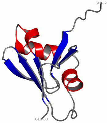
 Visualization

(-) Interactive Views

NMR Structure
  Complete Structure
    Jena3D(integrated viewing of ligand, site, SAP, PROSITE, SCOP information)
    WebMol | AstexViewer[tm]@PDBe
(Java Applets, require no local installation except for Java; loading may be slow)
    STRAP
(Java WebStart application, automatic local installation, requires Java; full application with system access!)
    RasMol
(require local installation)
    Molscript (VRML)
(requires installation of a VRML viewer; select preferred view via VRML and generate a mono or stereo PDF format file)
 
  Ligands, Modified Residues, Ions
(no "Ligands, Modified Residues, Ions" information available for 2uwq)
 
  Sites
(no "Sites" information available for 2uwq)
 
  Cis Peptide Bonds
(no "Cis Peptide Bonds" information available for 2uwq)
 

(-) Still Images

Jmol
  protein: cartoon or spacefill or dots and stick; nucleic acid: cartoon and stick; ligands: spacefill; active site: stick
Molscript
  protein, nucleic acid: cartoon; ligands: spacefill; active site: ball and stick

 Databases and Analysis Tools

(-) Databases

Access by PDB/NDB ID
  2uwq
    Family and Domain InformationProDom | SYSTERS
    General Structural InformationGlycoscienceDB | MMDB | NDB | OCA | PDB | PDBe | PDBj | PDBsum | PDBWiki | PQS | PROTEOPEDIA
    Orientation in MembranesOPM
    Protein SurfaceSURFACE
    Secondary StructureDSSP (structure derived) | HSSP (homology derived)
    Structural GenomicsGeneCensus
    Structural NeighboursCE | VAST
    Structure ClassificationCATH | Dali | SCOP
    Validation and Original DataBMRB Data View | BMRB Restraints Grid | EDS | PROCHECK | RECOORD | WHAT_CHECK
 
Access by UniProt ID/Accession number
  ASPP2_HUMAN | Q13625
    Comparative Protein Structure ModelsModBase
    Genomic InformationEnsembl
    Protein-protein InteractionDIP
    Sequence, Family and Domain InformationInterPro | Pfam | SMART | UniProtKB/SwissProt
 
Access by Enzyme Classificator   (EC Number)
  (no 'Enzyme Classificator' available)
    General Enzyme InformationBRENDA | EC-PDB | Enzyme | IntEnz
    PathwayKEGG | MetaCyc
 
Access by Disease Identifier   (MIM ID)
  (no 'MIM ID' available)
    Disease InformationOMIM
 
Access by GenAge ID
  (no 'GenAge ID' available)
    Age Related InformationGenAge

(-) Analysis Tools

Access by PDB/NDB ID
    Domain InformationXDom
    Interatomic Contacts of Structural UnitsCSU
    Ligand-protein ContactsLPC
    Protein CavitiescastP
    Sequence and Secondary StructurePDBCartoon
    Structure AlignmentSTRAP(Java WebStart application, automatic local installation, requires Java; full application with system access!)
    Structure and Sequence BrowserSTING
 
Access by UniProt ID/Accession number
  ASPP2_HUMAN | Q13625
    Protein Disorder PredictionDisEMBL | FoldIndex | GLOBPLOT (for more information see DisProt)

 Related Entries

(-) Entries Sharing at Least One Protein Chain (UniProt ID)

UniProtKB/Swiss-Prot
        ASPP2_HUMAN | Q136251ycs 4a63 4irv

(-) Related Entries Specified in the PDB File

1ycs P53-53BP2 COMPLEX