|
|
|
|
 Description
|
|
Compounds
|
||||||||||||||||||||||||||||||||||||||||||||||||||||
Chains, Units
Summary Information (see also Sequences/Alignments below) |
Ligands, Modified Residues, Ions (1, 1)
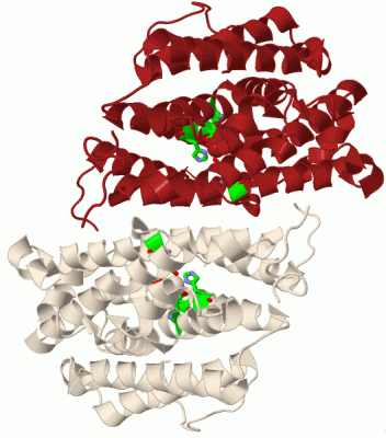
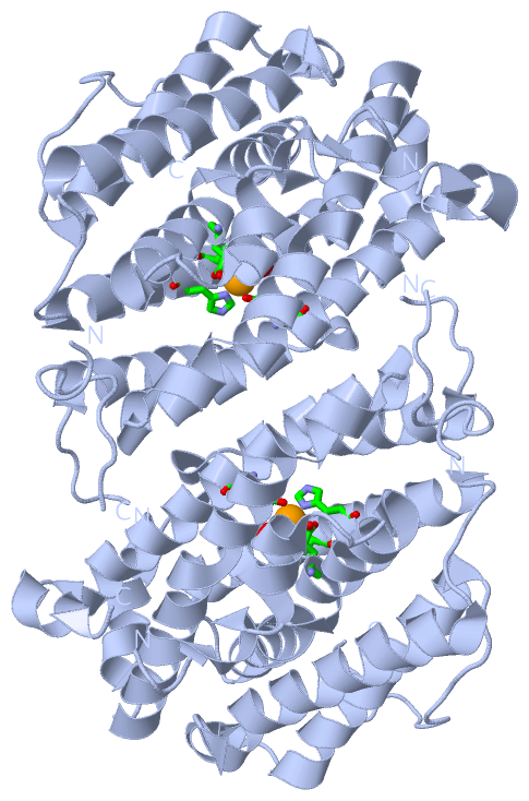
Asymmetric Unit (1, 1)
|
Sites (1, 1)
Asymmetric Unit (1, 1)
|
SS Bonds (0, 0)| (no "SS Bond" information available for 2UW2) |
Cis Peptide Bonds (1, 1)
Asymmetric Unit
|
||||||||
SAPs(SNPs)/Variants (0, 0)| (no "SAP(SNP)/Variant" information available for 2UW2) |
PROSITE Motifs (1, 1)
Asymmetric Unit (1, 1)
|
||||||||||||||||||||||||||||||||||||||||||||||||
Exons (8, 8)
Sequences/Alignments
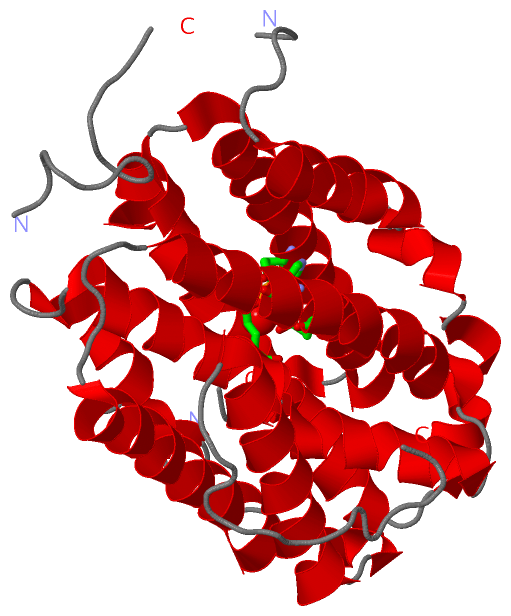
Asymmetric UnitChain A from PDB Type:PROTEIN Length:275 aligned with RIR2_HUMAN | P31350 from UniProtKB/Swiss-Prot Length:389 Alignment length:285 75 85 95 105 115 125 135 145 155 165 175 185 195 205 215 225 235 245 255 265 275 285 295 305 315 325 335 345 RIR2_HUMAN 66 GVEDEPLLRENPRRFVIFPIEYHDIWQMYKKAEASFWTAEEVDLSKDIQHWESLKPEERYFISHVLAFFAASDGIVNENLVERFSQEVQITEARCFYGFQIAMENIHSEMYSLLIDTYIKDPKEREFLFNAIETMPCVKKKADWALRWIGDKEATYGERVVAFAAVEGIFFSGSFASIFWLKKRGLMPGLTFSNELISRDEGLHCDFACLMFKHLVHKPSEERVREIIINAVRIEQEFLTEALPVKLIGMNCTLMKQYIEFVADRLMLELGFSKVFRVENPFDFM 350 SCOP domains d2uw2a1 A:66 -350 Ribonucleotide reductase R2 SCOP domains CATH domains --------------------------------------------------------------------------------------------------------------------------------------------------------------------------------------------------------------------------------------------------------------------------------------------- CATH domains Pfam domains ----Ribonuc_ red_sm-2uw2A01 A:70-350 Pfam domains SAPs(SNPs) --------------------------------------------------------------------------------------------------------------------------------------------------------------------------------------------------------------------------------------------------------------------------------------------- SAPs(SNPs) PROSITE ------------------------------------------------------------------------------------------------------RIBORED_SMALL ---------------------------------------------------------------------------------------------------------------------------------------------------------------------- PROSITE Transcript 1 (1) Exon 1.2c PDB: A:66-106 (gaps) Exon 1.3a PDB: A:107-145 Exon 1.4a PDB: A:146-190 UniProt: 146-190 -------------------------------Exon 1.6a PDB: A:222-266 UniProt: 222-266 Exon 1.7 PDB: A:267-301 Exon 1.8a PDB: A:302-339 Exon 1.9b Transcript 1 (1) Transcript 1 (2) ----------------------------------------------------------------------------------------------------------------------------Exon 1.5e PDB: A:190-222 (gaps) -------------------------------------------------------------------------------------------------------------------------------- Transcript 1 (2) 2uw2 A 66 GVEDEPLLRENP----IFPIEYHDIWQMYKKAEASFWTAEEVDLSKDIQHWESLKPEERYFISHVLAFFAASDGIVNENLVERFSQEVQITEARCFYGFQIAMENIHSEMYSLLIDTYIKDPKEREFL------MPCVKKKADWALRWIGDKEATYGERVVAFAAVEGIFFSGSFASIFWLKKRGPMPGLTFSNELISRDEGLHCDFACLMFKHLVHKPSEERVREIIINAVRIEQEFLTEALPVKLIGMNCTLMKQYIEFVADRLMLELGFSKVFRVENPFDFM 350 75 | | 85 95 105 115 125 135 145 155 165 175 185 | - | 205 215 225 235 245 255 265 275 285 295 305 315 325 335 345 77 82 193 200
|
||||||||||||||||||||
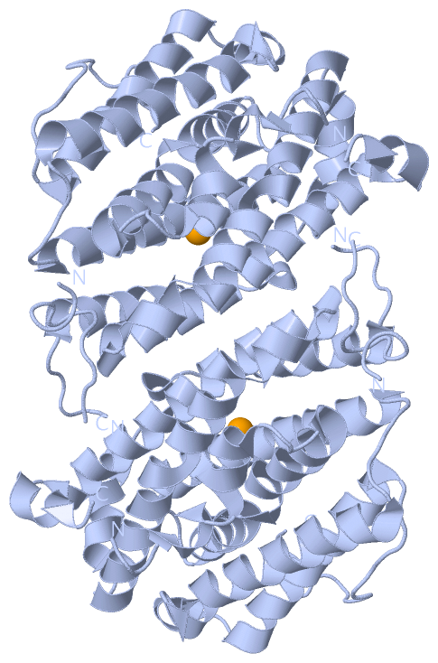
SCOP Domains (1, 1)
Asymmetric Unit

|
CATH Domains (0, 0 ; only for superseded entry 2IYH: 1,1)| (no "CATH Domain" information available for 2UW2, only for superseded entry 2IYH replaced by 2UW2) |
Pfam Domains (1, 1)
Asymmetric Unit
|
Gene Ontology (19, 19)|
Asymmetric Unit(hide GO term definitions) Chain A (RIR2_HUMAN | P31350)
|
||||||||||||||||||||||||||||||||||||||||||||||||||||||||||||||||||||||||||||||||||||||||||||||||||||||||||||||||||||||||||||||||||||
Interactive Views
|
|||||||||||||||||||||||||||||||||||||||||||||||||||||||||||||||||||||||||||||||||||||||||||||||||||||||||||||||||||||||||||||||||||||||||
Still Images
|
||||||||||||||||
Databases
|
||||||||||||||||||||||||||||||||||||||||||||||||||||||||||||||||||||||||||||||||||||||||||||||||||||||||||||||||||||||||||||||||||||||||||||||||||||||||||||||||
Analysis Tools
|
|||||||||||||||||||||||||||||||||||||||||||||||||||||||||||||
Entries Sharing at Least One Protein Chain (UniProt ID)
Related Entries Specified in the PDB File
|
|