Show PDB file:   
         Plain Text   HTML   (compressed file size)
QuickSearch:   
by PDB,NDB,UniProt,PROSITE Code or Search Term(s)  
(-)NMR Structure - model 1
(-)NMR Structure - all models
collapse expand < >
Image NMR Structure - model 1
NMR Structure - model 1  (Jmol Viewer)
Image NMR Structure - all models
NMR Structure - all models  (Jmol Viewer)

(-) Description

Title :  SOLUTION STRUCTURE OF THE CHROMODOMAIN OF HP1A WITH THE PHOSPHORYLATED N-TERMINAL TAIL COMPLEXED WITH H3K9ME3 PEPTIDE
 
Authors :  A. Kawaguchi, Y. Nishimura
Date :  18 Dec 15  (Deposition) - 16 Mar 16  (Release) - 16 Mar 16  (Revision)
Method :  SOLUTION NMR
Resolution :  NOT APPLICABLE
Chains :  NMR Structure  :  A,B  (20x)
NMR Structure *:  A,B  (1x)
Keywords :  Chromodomain, Hp1Alpha, Transcription-Peptide Complex (Keyword Search: [Gene Ontology, PubMed, Web (Google))
 
Reference :  H. Shimojo, A. Kawaguchi, T. Oda, N. Hashiguchi, S. Omori, K. Moritsugu, A. Kidera, K. Hiragami-Hamada, J. Nakayama, M. Sato, Y. Nishimura
Extended String-Like Binding Of The Phosphorylated Hp1 Alph N-Terminal Tail To The Lysine 9-Methylated Histone H3 Tail
Sci Rep V. 6 22527 2016
PubMed-ID: 26934956  |  Reference-DOI: 10.1038/SREP22527

(-) Compounds

Molecule 1 - CHROMOBOX PROTEIN HOMOLOG 5
    ChainsA
    EngineeredYES
    Expression SystemESCHERICHIA COLI
    Expression System Taxid562
    Expression System VectorPCOLD
    Expression System Vector TypeVECTOR
    FragmentN-TERMINAL DOMAIN, UNP RESIDUES 1-80
    GeneCBX5, HP1A
    Organism CommonMOUSE
    Organism ScientificMUS MUSCULUS
    Organism Taxid10090
    Other DetailsFOR PHOSPHORYLATION OF SER THE EXPRESSION WITH PRSFDUET (CK2)
    SynonymHETEROCHROMATIN PROTEIN 1 HOMOLOG ALPHA, HP1 ALPHA
 
Molecule 2 - 18-MER PEPTIDE OF HISTONE H3
    ChainsB
    EngineeredYES
    Organism CommonMOUSE
    Organism ScientificMUS MUSCULUS
    Organism Taxid10090
    Other DetailsSYNTHETIC PEPTIDE
    SyntheticYES

 Structural Features

(-) Chains, Units

  12
NMR Structure (20x)AB
NMR Structure * (1x)AB

Summary Information (see also Sequences/Alignments below)
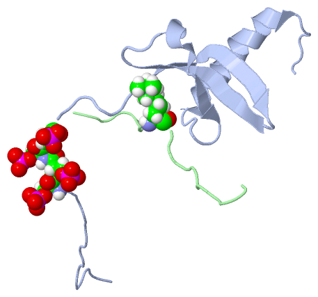
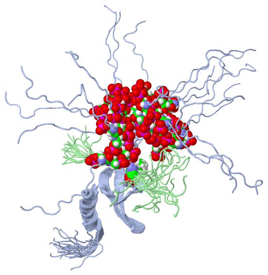
(-) Ligands, Modified Residues, Ions  (2, 5)

NMR Structure (2, 5)
No.NameCountTypeFull Name
1M3L1Mod. Amino AcidN-TRIMETHYLLYSINE
2SEP4Mod. Amino AcidPHOSPHOSERINE
NMR Structure * (2, 5)
No.NameCountTypeFull Name
1M3L1Mod. Amino AcidN-TRIMETHYLLYSINE
2SEP4Mod. Amino AcidPHOSPHOSERINE

(-) Sites  (0, 0)

(no "Site" information available for 2RVN)

(-) SS Bonds  (0, 0)

(no "SS Bond" information available for 2RVN)

(-) Cis Peptide Bonds  (0, 0)

(no "Cis Peptide Bond" information available for 2RVN)

 Sequence-Structure Mapping

(-) SAPs(SNPs)/Variants  (0, 0)

(no "SAP(SNP)/Variant" information available for 2RVN)

(-) PROSITE Motifs  (0, 0)

(no "PROSITE Motif" information available for 2RVN)

(-) Exons   (0, 0)

(no "Exon" information available for 2RVN)

(-) Sequences/Alignments

NMR Structure
   Reformat: Number of residues per line =  ('0' or empty: single-line sequence representation)
  Number of residues per labelling interval =   
  UniProt sequence: complete  aligned part    
   Show mapping: SCOP domains CATH domains Pfam domains Secondary structure (by author)
SAPs(SNPs) PROSITE motifs Exons
(details for a mapped element are shown in a popup box when the mouse pointer rests over it)
Chain A from PDB  Type:PROTEIN  Length:83
                                                                                                                  
               SCOP domains ----------------------------------------------------------------------------------- SCOP domains
               CATH domains ----------------------------------------------------------------------------------- CATH domains
               Pfam domains ----------------------------------------------------------------------------------- Pfam domains
         Sec.struct. author ........................eeeeeeeeee..eeeeeeee...hhhhh.eee.....hhhhhhhhhhhhhhhh...... Sec.struct. author
                 SAPs(SNPs) ----------------------------------------------------------------------------------- SAPs(SNPs)
                    PROSITE ----------------------------------------------------------------------------------- PROSITE
                 Transcript ----------------------------------------------------------------------------------- Transcript
                  2rvn A -2 GSHMGKKTKRTADssssEDEEEYVVEKVLDRRMVKGQVEYLLKWKGFSEEHNTWEPEKNLDCPELISEFMKKYKKMKEGENNK 80
                                     7   |||| 17        27        37        47        57        67        77   
                                        11-SEP                                                                 
                                         12-SEP                                                                
                                          13-SEP                                                               
                                           14-SEP                                                              

Chain B from PDB  Type:PROTEIN  Length:18
                                                 
               SCOP domains ------------------ SCOP domains
               CATH domains ------------------ CATH domains
               Pfam domains ------------------ Pfam domains
         Sec.struct. author .................. Sec.struct. author
                 SAPs(SNPs) ------------------ SAPs(SNPs)
                    PROSITE ------------------ PROSITE
                 Transcript ------------------ Transcript
                  2rvn B  1 ARTKQTARkSTGGKAPRY 18
                                    10        
                                    9-M3L     

   Legend:   → Mismatch (orange background)
  - → Gap (green background, '-', border residues have a numbering label)
    → Modified Residue (blue background, lower-case, 'x' indicates undefined single-letter code, labelled with number + name)
  x → Chemical Group (purple background, 'x', labelled with number + name, e.g. ACE or NH2)
  extra numbering lines below/above indicate numbering irregularities and modified residue names etc., number ends below/above '|'

 Classification and Annotation

(-) SCOP Domains  (0, 0)

(no "SCOP Domain" information available for 2RVN)

(-) CATH Domains  (0, 0)

(no "CATH Domain" information available for 2RVN)

(-) Pfam Domains  (0, 0)

(no "Pfam Domain" information available for 2RVN)

(-) Gene Ontology  (53, 56)

NMR Structure(hide GO term definitions)

 Visualization

(-) Interactive Views

NMR Structure
  Complete Structure
    Jena3D(integrated viewing of ligand, site, SAP, PROSITE, SCOP information)
    WebMol | AstexViewer[tm]@PDBe
(Java Applets, require no local installation except for Java; loading may be slow)
    STRAP
(Java WebStart application, automatic local installation, requires Java; full application with system access!)
    RasMol
(require local installation)
    Molscript (VRML)
(requires installation of a VRML viewer; select preferred view via VRML and generate a mono or stereo PDF format file)
 
  Ligands, Modified Residues, Ions
    M3L  [ RasMol | Jena3D ]  +environment [ RasMol | Jena3D ]
    SEP  [ RasMol | Jena3D ]  +environment [ RasMol | Jena3D ]
 
  Sites
(no "Sites" information available for 2rvn)
 
  Cis Peptide Bonds
(no "Cis Peptide Bonds" information available for 2rvn)
 

(-) Still Images

Jmol
  protein: cartoon or spacefill or dots and stick; nucleic acid: cartoon and stick; ligands: spacefill; active site: stick
Molscript
  protein, nucleic acid: cartoon; ligands: spacefill; active site: ball and stick

 Databases and Analysis Tools

(-) Databases

Access by PDB/NDB ID
  2rvn
    Family and Domain InformationProDom | SYSTERS
    General Structural InformationGlycoscienceDB | MMDB | NDB | OCA | PDB | PDBe | PDBj | PDBsum | PDBWiki | PQS | PROTEOPEDIA
    Orientation in MembranesOPM
    Protein SurfaceSURFACE
    Secondary StructureDSSP (structure derived) | HSSP (homology derived)
    Structural GenomicsGeneCensus
    Structural NeighboursCE | VAST
    Structure ClassificationCATH | Dali | SCOP
    Validation and Original DataBMRB Data View | BMRB Restraints Grid | EDS | PROCHECK | RECOORD | WHAT_CHECK
 
Access by UniProt ID/Accession number
  CBX5_MOUSE | Q61686
    Comparative Protein Structure ModelsModBase
    Genomic InformationEnsembl
    Protein-protein InteractionDIP
    Sequence, Family and Domain InformationInterPro | Pfam | SMART | UniProtKB/SwissProt
  H33_MOUSE | P84244
    Comparative Protein Structure ModelsModBase
    Genomic InformationEnsembl
    Protein-protein InteractionDIP
    Sequence, Family and Domain InformationInterPro | Pfam | SMART | UniProtKB/SwissProt
 
Access by Enzyme Classificator   (EC Number)
  (no 'Enzyme Classificator' available)
    General Enzyme InformationBRENDA | EC-PDB | Enzyme | IntEnz
    PathwayKEGG | MetaCyc
 
Access by Disease Identifier   (MIM ID)
  (no 'MIM ID' available)
    Disease InformationOMIM
 
Access by GenAge ID
  (no 'GenAge ID' available)
    Age Related InformationGenAge

(-) Analysis Tools

Access by PDB/NDB ID
    Domain InformationXDom
    Interatomic Contacts of Structural UnitsCSU
    Ligand-protein ContactsLPC
    Protein CavitiescastP
    Sequence and Secondary StructurePDBCartoon
    Structure AlignmentSTRAP(Java WebStart application, automatic local installation, requires Java; full application with system access!)
    Structure and Sequence BrowserSTING
 
Access by UniProt ID/Accession number
  CBX5_MOUSE | Q61686
    Protein Disorder PredictionDisEMBL | FoldIndex | GLOBPLOT (for more information see DisProt)
  H33_MOUSE | P84244
    Protein Disorder PredictionDisEMBL | FoldIndex | GLOBPLOT (for more information see DisProt)

 Related Entries

(-) Entries Sharing at Least One Protein Chain (UniProt ID)

UniProtKB/Swiss-Prot
        CBX5_MOUSE | Q616862rvl 2rvm
        H33_MOUSE | P842441pdq

(-) Related Entries Specified in the PDB File

2rvl 2rvm