Show PDB file:   
         Plain Text   HTML   (compressed file size)
QuickSearch:   
by PDB,NDB,UniProt,PROSITE Code or Search Term(s)  
(-)NMR Structure - model 1
(-)NMR Structure - model 1, sites
(-)NMR Structure - all models
collapse expand < >
Image NMR Structure - model 1
NMR Structure - model 1  (Jmol Viewer)
Image NMR Structure - model 1, sites
NMR Structure - model 1, sites  (Jmol Viewer)
Image NMR Structure - all models
NMR Structure - all models  (Jmol Viewer)

(-) Description

Title :  SOLUTION STRUCTURE OF THE PLANT HOMEODOMAIN (PHD) OF THE E3 SUMO LIGASE SIZ1 FROM RICE
 
Authors :  H. Shindo, W. Tsuchiya, R. Suzuki, T. Yamazaki
Date :  12 Jan 12  (Deposition) - 15 Aug 12  (Release) - 15 Aug 12  (Revision)
Method :  SOLUTION NMR
Resolution :  NOT APPLICABLE
Chains :  NMR Structure  :  A  (20x)
NMR Structure *:  A  (1x)
Keywords :  E3 Sumo Ligase, Plant Homeodomain (Phd), Histone Binding, Ligase (Keyword Search: [Gene Ontology, PubMed, Web (Google))
 
Reference :  H. Shindo, R. Suzuki, W. Tsuchiya, M. Taichi, Y. Nishiuchi, T. Yamazak
Phd Finger Of The Sumo Ligase Siz/Pias Family In Rice Reveals Specific Binding For Methylated Histone H3 At Lysin 4 And Arginine 2
Febs Lett. V. 586 1783 2012
PubMed-ID: 22626555  |  Reference-DOI: 10.1016/J.FEBSLET.2012.04.063

(-) Compounds

Molecule 1 - E3 SUMO-PROTEIN LIGASE SIZ1
    ChainsA
    EC Number6.3.2.-
    EngineeredYES
    Expression SystemESCHERICHIA COLI
    Expression System StrainBL21(DE3)
    Expression System Taxid562
    Expression System VectorPGEX-4T-3
    Expression System Vector TypeVECTOR
    FragmentPLANT HOMEODOMAIN, UNP RESIDUES 107-172
    GeneSIZ1, OS05G0125000, LOC_OS05G03430, OSJNBB0079L11.3
    Organism CommonJAPANESE RICE
    Organism ScientificORYZA SATIVA JAPONICA GROUP
    Organism Taxid39947

 Structural Features

(-) Chains, Units

  1
NMR Structure (20x)A
NMR Structure * (1x)A

Summary Information (see also Sequences/Alignments below)

(-) Ligands, Modified Residues, Ions  (1, 2)

NMR Structure (1, 2)
No.NameCountTypeFull Name
1ZN2Ligand/IonZINC ION
NMR Structure * (0, 0)
No.NameCountTypeFull Name
1ZN-1Ligand/IonZINC ION

(-) Sites  (2, 2)

NMR Structure (2, 2)
No.NameEvidenceResiduesDescription
1AC1SOFTWARECYS A:117 , CYS A:119 , HIS A:141 , CYS A:144BINDING SITE FOR RESIDUE ZN A 901
2AC2SOFTWARECYS A:131 , CYS A:136 , VAL A:138 , CYS A:163 , CYS A:166BINDING SITE FOR RESIDUE ZN A 902

(-) SS Bonds  (0, 0)

(no "SS Bond" information available for 2RSD)

(-) Cis Peptide Bonds  (0, 0)

(no "Cis Peptide Bond" information available for 2RSD)

 Sequence-Structure Mapping

(-) SAPs(SNPs)/Variants  (0, 0)

(no "SAP(SNP)/Variant" information available for 2RSD)

(-) PROSITE Motifs  (1, 1)

NMR Structure (1, 1)
 PROSITEUniProtKBPDB
No.IDACDescriptionIDLocationCountLocation
1ZF_PHD_1PS01359 Zinc finger PHD-type signature.SIZ1_ORYSJ117-166  1A:117-166
NMR Structure * (1, 1)
 PROSITEUniProtKBPDB
No.IDACDescriptionIDLocationCountLocation
1ZF_PHD_1PS01359 Zinc finger PHD-type signature.SIZ1_ORYSJ117-166  1A:117-166

(-) Exons   (0, 0)

(no "Exon" information available for 2RSD)

(-) Sequences/Alignments

NMR Structure
   Reformat: Number of residues per line =  ('0' or empty: single-line sequence representation)
  Number of residues per labelling interval =   
  UniProt sequence: complete  aligned part    
   Show mapping: SCOP domains CATH domains Pfam domains Secondary structure (by author)
SAPs(SNPs) PROSITE motifs Exons
(details for a mapped element are shown in a popup box when the mouse pointer rests over it)
Chain A from PDB  Type:PROTEIN  Length:68
 aligned with SIZ1_ORYSJ | Q6L4L4 from UniProtKB/Swiss-Prot  Length:875

    Alignment length:79
                                   103       113       123       133       143       153       163         
           SIZ1_ORYSJ    94 GSDFSFRPIEEAYDSFQPEAKVRCICSSTMVNDSMIQCEDQRCQVWQHLNCVLIPDKPGESAEVPPVFYCELCRLSRAD 172
               SCOP domains d2           rsda_ A: automated matches                                         SCOP domains
               CATH domains ------------------------------------------------------------------------------- CATH domains
               Pfam domains ------------------------------------------------------------------------------- Pfam domains
         Sec.struct. author ..-----------.......ee............eee.......eeee.....................hhhhhhhhh. Sec.struct. author
                 SAPs(SNPs) ------------------------------------------------------------------------------- SAPs(SNPs)
                    PROSITE -----------------------ZF_PHD_1  PDB: A:117-166 UniProt: 117-166         ------ PROSITE
                 Transcript ------------------------------------------------------------------------------- Transcript
                 2rsd A 105 GS-----------DSFQPEAKVRCICSSTMVNDSMIQCEDQRCQVWQHLNCVLIPDKPGESAEVPPVFYCELCRLSRAD 172
                             |       -   |   113       123       133       143       153       163         
                             |         107                                                                 
                           106                                                                             

   Legend:   → Mismatch (orange background)
  - → Gap (green background, '-', border residues have a numbering label)
    → Modified Residue (blue background, lower-case, 'x' indicates undefined single-letter code, labelled with number + name)
  x → Chemical Group (purple background, 'x', labelled with number + name, e.g. ACE or NH2)
  extra numbering lines below/above indicate numbering irregularities and modified residue names etc., number ends below/above '|'

 Classification and Annotation

(-) SCOP Domains  (1, 1)

NMR Structure

(-) CATH Domains  (0, 0)

(no "CATH Domain" information available for 2RSD)

(-) Pfam Domains  (0, 0)

(no "Pfam Domain" information available for 2RSD)

(-) Gene Ontology  (10, 10)

NMR Structure(hide GO term definitions)
Chain A   (SIZ1_ORYSJ | Q6L4L4)
molecular function
    GO:0061665    SUMO ligase activity    Catalysis of the transfer of SUMO to a substrate protein via the reaction X-SUMO + S --> X + S-SUMO, where X is either an E2 or E3 enzyme, the X-SUMO linkage is a thioester bond, and the S-SUMO linkage is an isopeptide bond between the C-terminal amino acid of SUMO and the epsilon-amino group of lysine residues in the substrate.
    GO:0019789    SUMO transferase activity    Catalysis of the transfer of SUMO from one protein to another via the reaction X-SUMO + Y --> Y-SUMO + X, where both X-SUMO and Y-SUMO are covalent linkages.
    GO:0016874    ligase activity    Catalysis of the joining of two substances, or two groups within a single molecule, with the concomitant hydrolysis of the diphosphate bond in ATP or a similar triphosphate.
    GO:0046872    metal ion binding    Interacting selectively and non-covalently with any metal ion.
    GO:0005515    protein binding    Interacting selectively and non-covalently with any protein or protein complex (a complex of two or more proteins that may include other nonprotein molecules).
    GO:0008270    zinc ion binding    Interacting selectively and non-covalently with zinc (Zn) ions.
biological process
    GO:0016049    cell growth    The process in which a cell irreversibly increases in size over time by accretion and biosynthetic production of matter similar to that already present.
    GO:0031668    cellular response to extracellular stimulus    Any process that results in a change in state or activity of a cell (in terms of movement, secretion, enzyme production, gene expression, etc.) as a result of an extracellular stimulus.
    GO:0016925    protein sumoylation    The process in which a SUMO protein (small ubiquitin-related modifier) is conjugated to a target protein via an isopeptide bond between the carboxyl terminus of SUMO with an epsilon-amino group of a lysine residue of the target protein.
cellular component
    GO:0005634    nucleus    A membrane-bounded organelle of eukaryotic cells in which chromosomes are housed and replicated. In most cells, the nucleus contains all of the cell's chromosomes except the organellar chromosomes, and is the site of RNA synthesis and processing. In some species, or in specialized cell types, RNA metabolism or DNA replication may be absent.
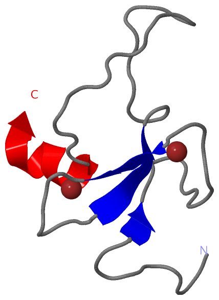
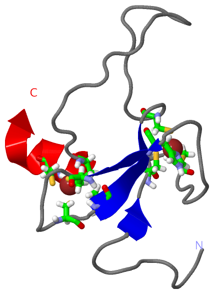
 Visualization

(-) Interactive Views

NMR Structure
  Complete Structure
    Jena3D(integrated viewing of ligand, site, SAP, PROSITE, SCOP information)
    WebMol | AstexViewer[tm]@PDBe
(Java Applets, require no local installation except for Java; loading may be slow)
    STRAP
(Java WebStart application, automatic local installation, requires Java; full application with system access!)
    RasMol
(require local installation)
    Molscript (VRML)
(requires installation of a VRML viewer; select preferred view via VRML and generate a mono or stereo PDF format file)
 
  Ligands, Modified Residues, Ions
    ZN  [ RasMol | Jena3D ]  +environment [ RasMol | Jena3D ]
 
  Sites
    AC1  [ RasMol ]  +environment [ RasMol ]
    AC2  [ RasMol ]  +environment [ RasMol ]
 
  Cis Peptide Bonds
(no "Cis Peptide Bonds" information available for 2rsd)
 

(-) Still Images

Jmol
  protein: cartoon or spacefill or dots and stick; nucleic acid: cartoon and stick; ligands: spacefill; active site: stick
Molscript
  protein, nucleic acid: cartoon; ligands: spacefill; active site: ball and stick

 Databases and Analysis Tools

(-) Databases

Access by PDB/NDB ID
  2rsd
    Family and Domain InformationProDom | SYSTERS
    General Structural InformationGlycoscienceDB | MMDB | NDB | OCA | PDB | PDBe | PDBj | PDBsum | PDBWiki | PQS | PROTEOPEDIA
    Orientation in MembranesOPM
    Protein SurfaceSURFACE
    Secondary StructureDSSP (structure derived) | HSSP (homology derived)
    Structural GenomicsGeneCensus
    Structural NeighboursCE | VAST
    Structure ClassificationCATH | Dali | SCOP
    Validation and Original DataBMRB Data View | BMRB Restraints Grid | EDS | PROCHECK | RECOORD | WHAT_CHECK
 
Access by UniProt ID/Accession number
  SIZ1_ORYSJ | Q6L4L4
    Comparative Protein Structure ModelsModBase
    Genomic InformationEnsembl
    Protein-protein InteractionDIP
    Sequence, Family and Domain InformationInterPro | Pfam | SMART | UniProtKB/SwissProt
 
Access by Enzyme Classificator   (EC Number)
  6.3.2.-
    General Enzyme InformationBRENDA | EC-PDB | Enzyme | IntEnz
    PathwayKEGG | MetaCyc
 
Access by Disease Identifier   (MIM ID)
  (no 'MIM ID' available)
    Disease InformationOMIM
 
Access by GenAge ID
  (no 'GenAge ID' available)
    Age Related InformationGenAge

(-) Analysis Tools

Access by PDB/NDB ID
    Domain InformationXDom
    Interatomic Contacts of Structural UnitsCSU
    Ligand-protein ContactsLPC
    Protein CavitiescastP
    Sequence and Secondary StructurePDBCartoon
    Structure AlignmentSTRAP(Java WebStart application, automatic local installation, requires Java; full application with system access!)
    Structure and Sequence BrowserSTING
 
Access by UniProt ID/Accession number
  SIZ1_ORYSJ | Q6L4L4
    Protein Disorder PredictionDisEMBL | FoldIndex | GLOBPLOT (for more information see DisProt)

 Related Entries

(-) Entries Sharing at Least One Protein Chain (UniProt ID)

UniProtKB/Swiss-Prot
        SIZ1_ORYSJ | Q6L4L42rno

(-) Related Entries Specified in the PDB File

(no "Related Entries Specified in the PDB File" available for 2RSD)