|
|
|
|
 Description
|
|
Compounds
|
||||||||||||||||||||||||||||||||||||||||||||
Chains, Units
Summary Information (see also Sequences/Alignments below) |
Ligands, Modified Residues, Ions (0, 0)| (no "Ligand,Modified Residues,Ions" information available for 2RR7) |
Sites (0, 0)| (no "Site" information available for 2RR7) |
SS Bonds (0, 0)| (no "SS Bond" information available for 2RR7) |
Cis Peptide Bonds (0, 0)| (no "Cis Peptide Bond" information available for 2RR7) |
SAPs(SNPs)/Variants (0, 0)| (no "SAP(SNP)/Variant" information available for 2RR7) |
PROSITE Motifs (0, 0)| (no "PROSITE Motif" information available for 2RR7) |
Exons (0, 0)| (no "Exon" information available for 2RR7) |
Sequences/Alignments
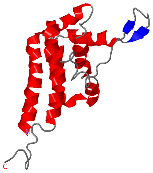
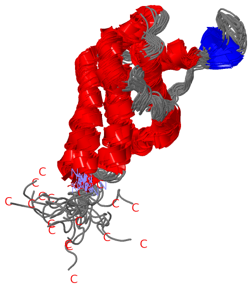
NMR StructureChain A from PDB Type:PROTEIN Length:155 aligned with Q4AC22_CHLRE | Q4AC22 from UniProtKB/TrEMBL Length:4149 Alignment length:155 2797 2807 2817 2827 2837 2847 2857 2867 2877 2887 2897 2907 2917 2927 2937 Q4AC22_CHLRE 2788 ECEADLAEALPLLEAALKALDTLKPADITEVKGMKSPPAGVRRVLEAICIMKGVKPARVKDTASGRMVDDYWEASKKMLMEFDFLDSLRKFDKDHIPPEVIVKIRPFAQDPEFQPKVIEKQSVACAGLCSWVIALEKYDKVIKEVEPKRQKLREA 2942 SCOP domains ----------------------------------------------------------------------------------------------------------------------------------------------------------- SCOP domains CATH domains ----------------------------------------------------------------------------------------------------------------------------------------------------------- CATH domains Pfam domains ---MT-2rr7A01 A:4-155 Pfam domains SAPs(SNPs) ----------------------------------------------------------------------------------------------------------------------------------------------------------- SAPs(SNPs) PROSITE ----------------------------------------------------------------------------------------------------------------------------------------------------------- PROSITE Transcript ----------------------------------------------------------------------------------------------------------------------------------------------------------- Transcript 2rr7 A 1 GSQADLAEALPLLEAALKALDTLKPADITEVKGMKSPPAGVRRVLEAICIMKGVKPARVKDTASGRMVDDYWEASKKMLMEFDFLDSLRKFDKDHIPPEVIVKIRPFAQDPEFQPKVIEKQSVACAGLCSWVIALEKYDKVIKEVEPKRQKLREA 155 10 20 30 40 50 60 70 80 90 100 110 120 130 140 150
|
||||||||||||||||||||
SCOP Domains (0, 0)| (no "SCOP Domain" information available for 2RR7) |
CATH Domains (0, 0)| (no "CATH Domain" information available for 2RR7) |
Pfam Domains (1, 1)
NMR Structure
|
Gene Ontology (4, 4)|
NMR Structure(hide GO term definitions) Chain A (Q4AC22_CHLRE | Q4AC22)
|
||||||||||||||||||||||||||||||||||||||||||
Interactive Views
|
||||||||||||||||||||||||||||||||||||||||||||||||||||||||||||||||||||||||||||||||||||||||||||||||||||||||||||||||||||
Still Images
|
||||||||||||||||
Databases
|
||||||||||||||||||||||||||||||||||||||||||||||||||||||||||||||||||||||||||||||||||||||||||||||||||||||||||||||||||||||||||||||||||||||||||||||||||||||||||||||||
Analysis Tools
|
|||||||||||||||||||||||||||||||||||||||||||||||||||||||||||||
Entries Sharing at Least One Protein Chain (UniProt ID)
Related Entries Specified in the PDB File
|
|