|
|
|
|
 Description
|
|
Compounds
|
||||||||||||||||||||||||||||||||||||||||
Chains, Units
Summary Information (see also Sequences/Alignments below) |
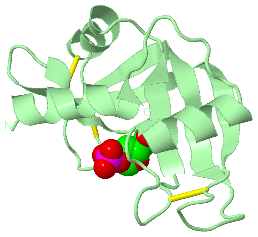
Ligands, Modified Residues, Ions (1, 2)
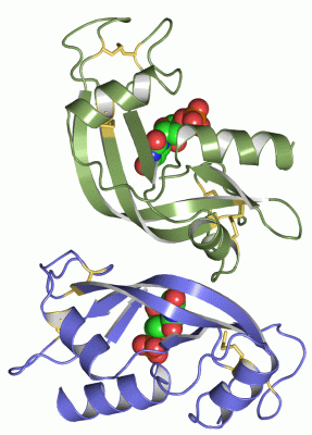
Asymmetric Unit (1, 2)
|
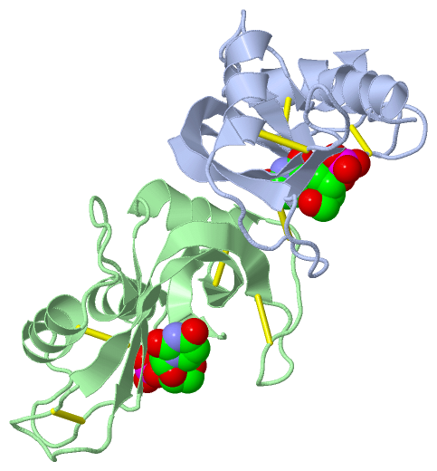
Sites (2, 2)
Asymmetric Unit (2, 2)
|
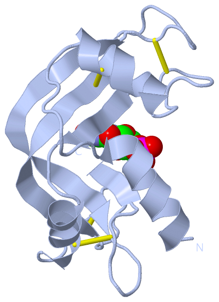
SS Bonds (8, 8)
Asymmetric Unit
|
||||||||||||||||||||||||||||||||||||
Cis Peptide Bonds (4, 4)
Asymmetric Unit
|
||||||||||||||||||||
SAPs(SNPs)/Variants (0, 0)| (no "SAP(SNP)/Variant" information available for 2RNF) |
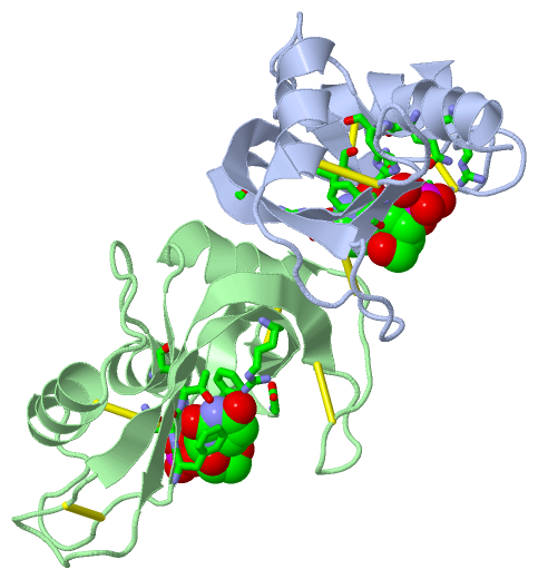
PROSITE Motifs (1, 2)
Asymmetric Unit (1, 2)
|
||||||||||||||||||||||||||||||||||||||||||||||||||||||||||||||||||||||||
Exons (0, 0)| (no "Exon" information available for 2RNF) |
Sequences/Alignments
Asymmetric UnitChain A from PDB Type:PROTEIN Length:120 aligned with RNAS4_HUMAN | P34096 from UniProtKB/Swiss-Prot Length:147 Alignment length:120 37 47 57 67 77 87 97 107 117 127 137 147 RNAS4_HUMAN 28 GQDGMYQRFLRQHVHPEETGGSDRYCNLMMQRRKMTLYHCKRFNTFIHEDIWNIRSICSTTNIQCKNGKMNCHEGVVKVTDCRDTGSSRAPNCRYRAIASTRRVVIACEGNPQVPVHFDG 147 SCOP domains d2rnfa_ A: Ribonuclease 4 SCOP domains CATH domains 2rnfA00 A:0-119 P-30 Protein CATH domains Pfam domains ------------------------------------------------------------------------------------------------------------------------ Pfam domains SAPs(SNPs) ------------------------------------------------------------------------------------------------------------------------ SAPs(SNPs) PROSITE ---------------------------------------RNASE_P-------------------------------------------------------------------------- PROSITE Transcript ------------------------------------------------------------------------------------------------------------------------ Transcript 2rnf A 0 MQDGMYQRFLRQHVHPEETGGSDRYCNLMMQRRKMTLYHCKRFNTFIHEDIWNIRSICSTTNIQCKNGKMNCHEGVVKVTDCRDTGSSRAPNCRYRAIASTRRVVIACEGNPQVPVHFDG 119 9 19 29 39 49 59 69 79 89 99 109 119 Chain B from PDB Type:PROTEIN Length:120 aligned with RNAS4_HUMAN | P34096 from UniProtKB/Swiss-Prot Length:147 Alignment length:120 37 47 57 67 77 87 97 107 117 127 137 147 RNAS4_HUMAN 28 GQDGMYQRFLRQHVHPEETGGSDRYCNLMMQRRKMTLYHCKRFNTFIHEDIWNIRSICSTTNIQCKNGKMNCHEGVVKVTDCRDTGSSRAPNCRYRAIASTRRVVIACEGNPQVPVHFDG 147 SCOP domains d2rnfb_ B: Ribonuclease 4 SCOP domains CATH domains 2rnfB00 B:0-119 P-30 Protein CATH domains Pfam domains (1) -RnaseA-2rnfB01 B:1-118 - Pfam domains (1) Pfam domains (2) -RnaseA-2rnfB02 B:1-118 - Pfam domains (2) SAPs(SNPs) ------------------------------------------------------------------------------------------------------------------------ SAPs(SNPs) PROSITE ---------------------------------------RNASE_P-------------------------------------------------------------------------- PROSITE Transcript ------------------------------------------------------------------------------------------------------------------------ Transcript 2rnf B 0 MQDGMYQRFLRQHVHPEETGGSDRYCNLMMQRRKMTLYHCKRFNTFIHEDIWNIRSICSTTNIQCKNGKMNCHEGVVKVTDCRDTGSSRAPNCRYRAIASTRRVVIACEGNPQVPVHFDG 119 9 19 29 39 49 59 69 79 89 99 109 119
|
||||||||||||||||||||
SCOP Domains (1, 2)
Asymmetric Unit
 |
CATH Domains (1, 2)
Asymmetric Unit
|
Pfam Domains (1, 2)
Asymmetric Unit
|
Gene Ontology (10, 10)|
Asymmetric Unit(hide GO term definitions) Chain A,B (RNAS4_HUMAN | P34096)
|
||||||||||||||||||||||||||||||||||||||||||||||||||||||||||||||||||||||||||||||
Interactive Views
|
||||||||||||||||||||||||||||||||||||||||||||||||||||||||||||||||||||||||||||||||||||||||||||||||||||||||||||||||||||||||||||||||||||||||||||||||||||||||||||||||||||||||||
Still Images
|
||||||||||||||||
Databases
|
||||||||||||||||||||||||||||||||||||||||||||||||||||||||||||||||||||||||||||||||||||||||||||||||||||||||||||||||||||||||||||||||||||||||||||||||||||||||||||||||
Analysis Tools
|
|||||||||||||||||||||||||||||||||||||||||||||||||||||||||||||
Entries Sharing at Least One Protein Chain (UniProt ID)
Related Entries Specified in the PDB File
|
|