|
|
|
|
 Description
|
|
Compounds
|
||||||||||||||||||||||||||||||||||||||||||||||||||||
Chains, Units
Summary Information (see also Sequences/Alignments below) |
Ligands, Modified Residues, Ions (0, 0)| (no "Ligand,Modified Residues,Ions" information available for 2RK5) |
Sites (0, 0)| (no "Site" information available for 2RK5) |
SS Bonds (0, 0)| (no "SS Bond" information available for 2RK5) |
Cis Peptide Bonds (0, 0)| (no "Cis Peptide Bond" information available for 2RK5) |
SAPs(SNPs)/Variants (0, 0)| (no "SAP(SNP)/Variant" information available for 2RK5) |
PROSITE Motifs (0, 0)| (no "PROSITE Motif" information available for 2RK5) |
Exons (0, 0)| (no "Exon" information available for 2RK5) |
Sequences/Alignments
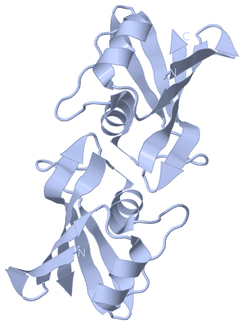
Asymmetric UnitChain A from PDB Type:PROTEIN Length:86 aligned with O68574_STRMG | O68574 from UniProtKB/TrEMBL Length:445 Alignment length:86 359 369 379 389 399 409 419 429 O68574_STRMG 350 SVREIADNTYIVLGTMTLNDFNEYFETDLESDNVDTIAGFYLTGVGTIPSQEEKEHFEVESNGKHLELINDKVKDGRVTKLKILVS 435 SCOP domains --d2rk5a1 A:5-88 Putative hemolysin SMU1693 SCOP domains CATH domains 2rk5A00 A:3-88 [code=3.30.465.10, no name defined] CATH domains Pfam domains -------------------------------------------------------------------------------------- Pfam domains SAPs(SNPs) -------------------------------------------------------------------------------------- SAPs(SNPs) PROSITE -------------------------------------------------------------------------------------- PROSITE Transcript -------------------------------------------------------------------------------------- Transcript 2rk5 A 3 QSREIADNTYIVLGTMTLNDFNEYFETDLESDNVDTIAGFYLTGVGTIPSQEEKEHFEVESNGKHLELINDKVKDGRVTKLKILVS 88 12 22 32 42 52 62 72 82
|
||||||||||||||||||||
SCOP Domains (1, 1)
Asymmetric Unit
 |
CATH Domains (1, 1)
Asymmetric Unit
|
Pfam Domains (0, 0)| (no "Pfam Domain" information available for 2RK5) |
Gene Ontology (6, 6)|
Asymmetric Unit(hide GO term definitions) Chain A (O68574_STRMG | O68574)
|
||||||||||||||||||||||||||||||||||||||||||||||||||||||
Interactive Views
|
||||||||||||||||||||||||||||||||||||||||||||||||||||||||||||||||||||||||||||||||||||||||||||||||||||||||||||||||||||||||||||||||||||||
Still Images
|
||||||||||||||||
Databases
|
||||||||||||||||||||||||||||||||||||||||||||||||||||||||||||||||||||||||||||||||||||||||||||||||||||||||||||||||||||||||||||||||||||||||||||||||||||||||||||||||
Analysis Tools
|
|||||||||||||||||||||||||||||||||||||||||||||||||||||||||||||
Entries Sharing at Least One Protein Chain (UniProt ID)
Related Entries Specified in the PDB File
|
|