|
|
|
|
 Description
|
|
Compounds
|
||||||||||||||||||||||||||||
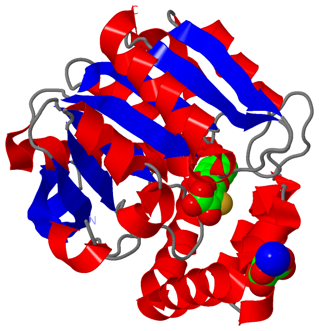
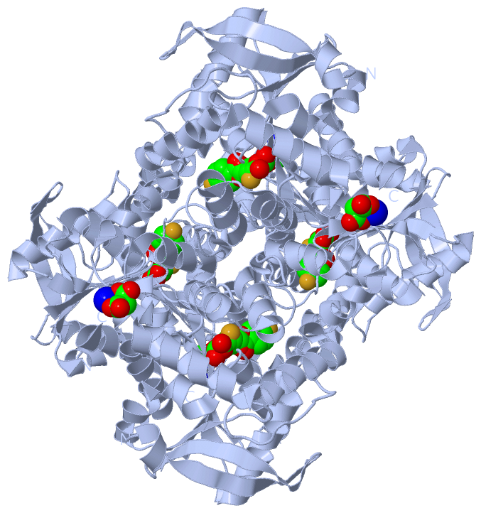
Chains, Units
Summary Information (see also Sequences/Alignments below) |
Ligands, Modified Residues, Ions (3, 4)| Asymmetric Unit (3, 4) Biological Unit 1 (2, 12) |
Sites (4, 4)
Asymmetric Unit (4, 4)
|
SS Bonds (0, 0)| (no "SS Bond" information available for 2RHW) |
Cis Peptide Bonds (1, 1)
Asymmetric Unit
|
||||||||
SAPs(SNPs)/Variants (0, 0)| (no "SAP(SNP)/Variant" information available for 2RHW) |
PROSITE Motifs (0, 0)| (no "PROSITE Motif" information available for 2RHW) |
Exons (0, 0)| (no "Exon" information available for 2RHW) |
Sequences/Alignments
Asymmetric UnitChain A from PDB Type:PROTEIN Length:283 aligned with BPHD_PARXL | P47229 from UniProtKB/Swiss-Prot Length:286 Alignment length:283 13 23 33 43 53 63 73 83 93 103 113 123 133 143 153 163 173 183 193 203 213 223 233 243 253 263 273 283 BPHD_PARXL 4 LTESSTSKFVKINEKGFSDFNIHYNEAGNGETVIMLHGGGPGAGGWSNYYRNVGPFVDAGYRVILKDSPGFNKSDAVVMDEQRGLVNARAVKGLMDALDIDRAHLVGNSMGGATALNFALEYPDRIGKLILMGPGGLGPSMFAPMPMEGIKLLFKLYAEPSYETLKQMLQVFLYDQSLITEELLQGRWEAIQRQPEHLKNFLISAQKAPLSTWDVTARLGEIKAKTFITWGRDDRFVPLDHGLKLLWNIDDARLHVFSKCGHWAQWEHADEFNRLVIDFLRHA 286 SCOP domains d2rhwa_ A: 2-hydroxy-6-oxo-6-phenylhexa-2,4-dienoate hydrolase (BPHD) SCOP domains CATH domains ------------------------------------------------------------------------------------------------------------------------------------------------------------------------------------------------------------------------------------------------------------------------------------------- CATH domains Pfam domains ------------------------------------------------------------------------------------------------------------------------------------------------------------------------------------------------------------------------------------------------------------------------------------------- Pfam domains SAPs(SNPs) ------------------------------------------------------------------------------------------------------------------------------------------------------------------------------------------------------------------------------------------------------------------------------------------- SAPs(SNPs) PROSITE ------------------------------------------------------------------------------------------------------------------------------------------------------------------------------------------------------------------------------------------------------------------------------------------- PROSITE Transcript ------------------------------------------------------------------------------------------------------------------------------------------------------------------------------------------------------------------------------------------------------------------------------------------- Transcript 2rhw A 4 LTESSTSKFVKINEKGFSDFNIHYNEAGNGETVIMLHGGGPGAGGWSNYYRNVGPFVDAGYRVILKDSPGFNKSDAVVMDEQRGLVNARAVKGLMDALDIDRAHLVGNAMGGATALNFALEYPDRIGKLILMGPGGLGPSMFAPMPMEGIKLLFKLYAEPSYETLKQMLQVFLYDQSLITEELLQGRWEAIQRQPEHLKNFLISAQKAPLSTWDVTARLGEIKAKTFITWGRDDRFVPLDHGLKLLWNIDDARLHVFSKCGHWAQWEHADEFNRLVIDFLRHA 286 13 23 33 43 53 63 73 83 93 103 113 123 133 143 153 163 173 183 193 203 213 223 233 243 253 263 273 283
|
||||||||||||||||||||
SCOP Domains (1, 1)
Asymmetric Unit
 |
CATH Domains (0, 0)| (no "CATH Domain" information available for 2RHW) |
Pfam Domains (0, 0)| (no "Pfam Domain" information available for 2RHW) |
Gene Ontology (6, 6)|
Asymmetric Unit(hide GO term definitions) Chain A (BPHD_PARXL | P47229)
|
||||||||||||||||||||||||||||||||||||||||||||||||
Interactive Views
|
||||||||||||||||||||||||||||||||||||||||||||||||||||||||||||||||||||||||||||||||||||||||||||||||||||||||||||||||||||||||||||||||||||||||||||||||||||||||||||||||||||||||||||
Still Images
|
||||||||||||||||
Databases
|
||||||||||||||||||||||||||||||||||||||||||||||||||||||||||||||||||||||||||||||||||||||||||||||||||||||||||||||||||||||||||||||||||||||||||||||||||||||||||||||||
Analysis Tools
|
|||||||||||||||||||||||||||||||||||||||||||||||||||||||||||||
Entries Sharing at Least One Protein Chain (UniProt ID)
Related Entries Specified in the PDB File
|
|