Show PDB file:   
         Plain Text   HTML   (compressed file size)
QuickSearch:   
by PDB,NDB,UniProt,PROSITE Code or Search Term(s)  
(-)Asym./Biol. Unit - manually
(-)Asym./Biol. Unit
(-)Asym./Biol. Unit - sites
collapse expand < >
Image Asym./Biol. Unit - manually
Asym./Biol. Unit - manually  (Jmol Viewer)
Image Asym./Biol. Unit
Asym./Biol. Unit  (Jmol Viewer)
Image Asym./Biol. Unit - sites
Asym./Biol. Unit - sites  (Jmol Viewer)

(-) Description

Title :  CRYSTAL STRUCTURE OF VCA0042 COMPLEXED WITH C-DI-GMP
 
Authors :  J. Benach, S. S. Swaminathan, R. Tamayo, J. Seetharaman, S. Handelman, F. Forouhar, H. Neely, A. Camilli, J. F. Hunt, Northeast Structural Consortium (Nesg)
Date :  22 Sep 07  (Deposition) - 30 Oct 07  (Release) - 13 Jul 11  (Revision)
Method :  X-RAY DIFFRACTION
Resolution :  1.92
Chains :  Asym./Biol. Unit :  A,B
Keywords :  C-Di-Gmp, Vibrio Cholerae, Vca0042, Structural Genomics, Psi-2, Protein Structure Initiative, Northeast Structural Genomics Consortium, Nesg, Unknown Function (Keyword Search: [Gene Ontology, PubMed, Web (Google))
 
Reference :  J. Benach, S. S. Swaminathan, R. Tamayo, S. K. Handelman, E. Folta-Stogniew, J. E. Ramos, F. Forouhar, H. Neely, J. Seetharaman A. Camilli, J. F. Hunt
The Structural Basis Of Cyclic Diguanylate Signal Transduction By Pilz Domains.
Embo J. V. 26 5153 2007
PubMed-ID: 18034161  |  Reference-DOI: 10.1038/SJ.EMBOJ.7601918

(-) Compounds

Molecule 1 - UNCHARACTERIZED PROTEIN VCA0042
    ChainsA, B
    EngineeredYES
    Expression SystemESCHERICHIA COLI BL21(DE3)
    Expression System PlasmidPET28B
    Expression System StrainBL21(DE3)
    Expression System Taxid469008
    Expression System Vector TypePLASMID
    GeneVCA0042, VC0395_0091
    Organism ScientificVIBRIO CHOLERAE
    Organism Taxid345073
    StrainO395

 Structural Features

(-) Chains, Units

  12
Asymmetric/Biological Unit AB

Summary Information (see also Sequences/Alignments below)

(-) Ligands, Modified Residues, Ions  (1, 2)

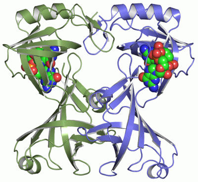
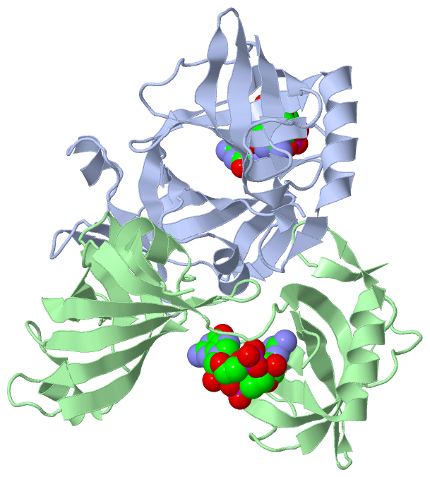
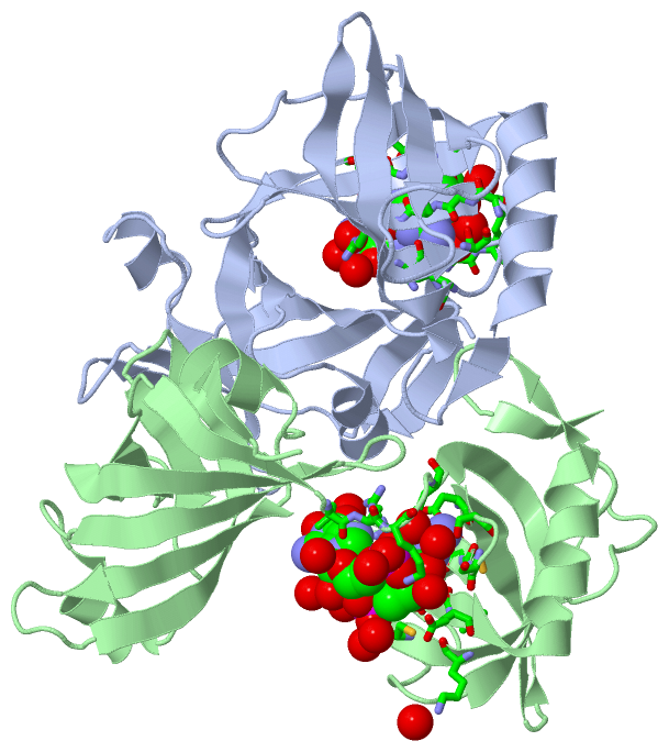
Asymmetric/Biological Unit (1, 2)
No.NameCountTypeFull Name
1C2E2Ligand/Ion9,9'-[(2R,3R,3AS,5S,7AR,9R,10R,10AS,12S,14AR)-3,5,10,12-TETRAHYDROXY-5,12-DIOXIDOOCTAHYDRO-2H,7H-DIFURO[3,2-D:3',2'-J][1,3,7,9,2,8]TETRAOXADIPHOSPHACYCLODODECINE-2,9-DIYL]BIS(2-AMINO-1,9-DIHYDRO-6H-PURIN-6-ONE)

(-) Sites  (2, 2)

Asymmetric Unit (2, 2)
No.NameEvidenceResiduesDescription
1AC1SOFTWARELEU A:135 , ARG A:136 , LYS A:137 , ARG A:140 , ASP A:162 , SER A:164 , SER A:166 , GLY A:167 , CYS A:168 , ARG A:169 , CYS A:207 , ASN A:208 , GLY A:219 , LEU A:220 , GLU A:221 , HOH A:311 , HOH A:313 , HOH A:316 , HOH A:321 , HOH A:429 , HOH A:446 , HOH A:449 , HOH A:477 , HOH A:533 , HOH A:549 , LYS B:205 , HOH B:531BINDING SITE FOR RESIDUE C2E A 301
2AC2SOFTWARELEU B:135 , ARG B:136 , LYS B:137 , ARG B:140 , ASP B:162 , SER B:164 , SER B:166 , GLY B:167 , CYS B:168 , ARG B:169 , CYS B:207 , ASN B:208 , GLY B:219 , LEU B:220 , GLU B:221 , HOH B:378 , HOH B:384 , HOH B:387 , HOH B:394 , HOH B:422 , HOH B:423 , HOH B:430 , HOH B:446 , HOH B:448 , HOH B:494 , HOH B:551 , HOH B:623 , HOH B:633BINDING SITE FOR RESIDUE C2E B 301

(-) SS Bonds  (0, 0)

(no "SS Bond" information available for 2RDE)

(-) Cis Peptide Bonds  (2, 2)

Asymmetric/Biological Unit
No.Residues
1Glu A:117 -Pro A:118
2Glu B:117 -Pro B:118

 Sequence-Structure Mapping

(-) SAPs(SNPs)/Variants  (0, 0)

(no "SAP(SNP)/Variant" information available for 2RDE)

(-) PROSITE Motifs  (0, 0)

(no "PROSITE Motif" information available for 2RDE)

(-) Exons   (0, 0)

(no "Exon" information available for 2RDE)

(-) Sequences/Alignments

Asymmetric/Biological Unit
   Reformat: Number of residues per line =  ('0' or empty: single-line sequence representation)
  Number of residues per labelling interval =   
  UniProt sequence: complete  aligned part    
   Show mapping: SCOP domains CATH domains Pfam domains Secondary structure (by author)
SAPs(SNPs) PROSITE motifs Exons
(details for a mapped element are shown in a popup box when the mouse pointer rests over it)
Chain A from PDB  Type:PROTEIN  Length:224
 aligned with A0A0H3ADR8_V | A0A0H3ADR8 from UniProtKB/TrEMBL  Length:252

    Alignment length:224
                                    33        43        53        63        73        83        93       103       113       123       133       143       153       163       173       183       193       203       213       223       233       243    
         A0A0H3ADR8_V    24 VSTINSTDALAMVEHSSELTLSITTPVGTKFVCRTPFIGTHTDKFLLVEMPKISADDLQYFFQEGFWMNIRAISPRGEGALIHFRSQLMHILQEPVPMAFLSIPNTMQVSQLRKEPRFELNLAGKVLFDEHRGDCELRDLSRSGCRFITPPLGKTYQVGDLVALEIFSDLRGTKTFPPLTGKICNLQRSLHHARYGLEFNEEGRNNAKNLLAQLKFNGTKLTLN 247
               SCOP domains d2rdea2 A:24-137 Hypothetical protein VCA0042, N-terminal domain                                                  d2rdea1 A:138-247 Hypothetical protein VCA0042, C-terminal domain                                              SCOP domains
               CATH domains 2rdeA01 A:24-136 Electron Transport, Fmn-binding Protein; Chain A                                                2rdeA02 A:137-247 predicted glycosyltransferase like domains                                                    CATH domains
               Pfam domains -------------------------------------------------------------------------------------------------------------------------------------------------------------------------------------------------------------------------------- Pfam domains
         Sec.struct. author .eeeehhhhhhh.....eeeeeee.....eeeeeeeeeeee...eeeee......hhhhh......eeeeeeee.....eeeeeeeeeeeee.....eeee....eeeeee......eeeeeeeeeee..eeeeeeeeee...eeeeee...........eeeeee............eeeeeeeeee....eeeeeeehhhhhhhhhhhhhhhee....ee.. Sec.struct. author
                 SAPs(SNPs) -------------------------------------------------------------------------------------------------------------------------------------------------------------------------------------------------------------------------------- SAPs(SNPs)
                    PROSITE -------------------------------------------------------------------------------------------------------------------------------------------------------------------------------------------------------------------------------- PROSITE
                 Transcript -------------------------------------------------------------------------------------------------------------------------------------------------------------------------------------------------------------------------------- Transcript
                 2rde A  24 VSTINSTDALAMVEHSSELTLSITTPVGTKFVCRTPFIGTHTDKFLLVEMPKISADDLQYFFQEGFWMNIRAISPRGEGALIHFRSQLMHILQEPVPMAFLSIPNTMQVSQLRKEPRFELNLAGKVLFDEHRGDCELRDLSRSGCRFITPPLGKTYQVGDLVALEIFSDLRGTKTFPPLTGKICNLQRSLHHARYGLEFNEEGRNNAKNLLAQLKFNGTKLTLN 247
                                    33        43        53        63        73        83        93       103       113       123       133       143       153       163       173       183       193       203       213       223       233       243    

Chain B from PDB  Type:PROTEIN  Length:225
 aligned with A0A0H3ADR8_V | A0A0H3ADR8 from UniProtKB/TrEMBL  Length:252

    Alignment length:225
                                    32        42        52        62        72        82        92       102       112       122       132       142       152       162       172       182       192       202       212       222       232       242     
         A0A0H3ADR8_V    23 TVSTINSTDALAMVEHSSELTLSITTPVGTKFVCRTPFIGTHTDKFLLVEMPKISADDLQYFFQEGFWMNIRAISPRGEGALIHFRSQLMHILQEPVPMAFLSIPNTMQVSQLRKEPRFELNLAGKVLFDEHRGDCELRDLSRSGCRFITPPLGKTYQVGDLVALEIFSDLRGTKTFPPLTGKICNLQRSLHHARYGLEFNEEGRNNAKNLLAQLKFNGTKLTLN 247
               SCOP domains d2rdeb2 B:23-137 Hypothetical protein VCA0042, N-terminal domain                                                   d2rdeb1 B:138-247 Hypothetical protein VCA0042, C-terminal domain                                              SCOP domains
               CATH domains 2rdeB01 B:23-136 Electron Transport, Fmn-binding Protein; Chain A                                                 2rdeB02 B:137-247 predicted glycosyltransferase like domains                                                    CATH domains
               Pfam domains --------------------------------------------------------------------------------------------------------------------------------------------------------------------------------------------------------------------------------- Pfam domains
         Sec.struct. author ..eeeehhhhhh......eeeeeee.....eeeeeeeeeeee...eeeee....hhhhhhhhh....eeeeeeee.....eeeeeeeeeeeee.....eeeee...eeeee.......eeeeeeeeeee..eeeeeeeeee...eeeeee...........eeeeee............eeeeeeeeee....eeeeeeehhhhhhhhhhhhh.eee....eee. Sec.struct. author
                 SAPs(SNPs) --------------------------------------------------------------------------------------------------------------------------------------------------------------------------------------------------------------------------------- SAPs(SNPs)
                    PROSITE --------------------------------------------------------------------------------------------------------------------------------------------------------------------------------------------------------------------------------- PROSITE
                 Transcript --------------------------------------------------------------------------------------------------------------------------------------------------------------------------------------------------------------------------------- Transcript
                 2rde B  23 TVSTINSTDALAMVEHSSELTLSITTPVGTKFVCRTPFIGTHTDKFLLVEMPKISADDLQYFFQEGFWMNIRAISPRGEGALIHFRSQLMHILQEPVPMAFLSIPNTMQVSQLRKEPRFELNLAGKVLFDEHRGDCELRDLSRSGCRFITPPLGKTYQVGDLVALEIFSDLRGTKTFPPLTGKICNLQRSLHHARYGLEFNEEGRNNAKNLLAQLKFNGTKLTLN 247
                                    32        42        52        62        72        82        92       102       112       122       132       142       152       162       172       182       192       202       212       222       232       242     

   Legend:   → Mismatch (orange background)
  - → Gap (green background, '-', border residues have a numbering label)
    → Modified Residue (blue background, lower-case, 'x' indicates undefined single-letter code, labelled with number + name)
  x → Chemical Group (purple background, 'x', labelled with number + name, e.g. ACE or NH2)
  extra numbering lines below/above indicate numbering irregularities and modified residue names etc., number ends below/above '|'

 Classification and Annotation

(-) SCOP Domains  (2, 4)

Asymmetric/Biological Unit

(-) CATH Domains  (2, 4)

Asymmetric/Biological Unit
(-)
Class: Mainly Beta (13760)

(-) Pfam Domains  (0, 0)

(no "Pfam Domain" information available for 2RDE)

(-) Gene Ontology  (0, 0)

Asymmetric/Biological Unit(hide GO term definitions)
    (no "Gene Ontology" information available for 2RDE)

 Visualization

(-) Interactive Views

Asymmetric/Biological Unit
  Complete Structure
    Jena3D(integrated viewing of ligand, site, SAP, PROSITE, SCOP information)
    WebMol | AstexViewer[tm]@PDBe
(Java Applets, require no local installation except for Java; loading may be slow)
    STRAP
(Java WebStart application, automatic local installation, requires Java; full application with system access!)
    RasMol
(require local installation)
    Molscript (VRML)
(requires installation of a VRML viewer; select preferred view via VRML and generate a mono or stereo PDF format file)
 
  Ligands, Modified Residues, Ions
    C2E  [ RasMol | Jena3D ]  +environment [ RasMol | Jena3D ]
 
  Sites
    AC1  [ RasMol ]  +environment [ RasMol ]
    AC2  [ RasMol ]  +environment [ RasMol ]
 
  Cis Peptide Bonds
    Glu A:117 - Pro A:118   [ RasMol ]  
    Glu B:117 - Pro B:118   [ RasMol ]  
 

(-) Still Images

Jmol
  protein: cartoon or spacefill or dots and stick; nucleic acid: cartoon and stick; ligands: spacefill; active site: stick
Molscript
  protein, nucleic acid: cartoon; ligands: spacefill; active site: ball and stick

 Databases and Analysis Tools

(-) Databases

Access by PDB/NDB ID
  2rde
    Family and Domain InformationProDom | SYSTERS
    General Structural InformationGlycoscienceDB | MMDB | NDB | OCA | PDB | PDBe | PDBj | PDBsum | PDBWiki | PQS | PROTEOPEDIA
    Orientation in MembranesOPM
    Protein SurfaceSURFACE
    Secondary StructureDSSP (structure derived) | HSSP (homology derived)
    Structural GenomicsGeneCensus
    Structural NeighboursCE | VAST
    Structure ClassificationCATH | Dali | SCOP
    Validation and Original DataBMRB Data View | BMRB Restraints Grid | EDS | PROCHECK | RECOORD | WHAT_CHECK
 
Access by UniProt ID/Accession number
  A0A0H3ADR8_V | A0A0H3ADR8
    Comparative Protein Structure ModelsModBase
    Genomic InformationEnsembl
    Protein-protein InteractionDIP
    Sequence, Family and Domain InformationInterPro | Pfam | SMART | UniProtKB/TrEMBL
 
Access by Enzyme Classificator   (EC Number)
  (no 'Enzyme Classificator' available)
    General Enzyme InformationBRENDA | EC-PDB | Enzyme | IntEnz
    PathwayKEGG | MetaCyc
 
Access by Disease Identifier   (MIM ID)
  (no 'MIM ID' available)
    Disease InformationOMIM
 
Access by GenAge ID
  (no 'GenAge ID' available)
    Age Related InformationGenAge

(-) Analysis Tools

Access by PDB/NDB ID
    Domain InformationXDom
    Interatomic Contacts of Structural UnitsCSU
    Ligand-protein ContactsLPC
    Protein CavitiescastP
    Sequence and Secondary StructurePDBCartoon
    Structure AlignmentSTRAP(Java WebStart application, automatic local installation, requires Java; full application with system access!)
    Structure and Sequence BrowserSTING
 
Access by UniProt ID/Accession number
  A0A0H3ADR8_V | A0A0H3ADR8
    Protein Disorder PredictionDisEMBL | FoldIndex | GLOBPLOT (for more information see DisProt)

 Related Entries

(-) Entries Sharing at Least One Protein Chain (UniProt ID)

(no "Entries Sharing at Least One Protein Chain" available for 2RDE)

(-) Related Entries Specified in the PDB File

(no "Related Entries Specified in the PDB File" available for 2RDE)