|
|
|
|
 Description
|
|
Compounds
|
||||||||||||||||||||||||||||||||||||||||||||||||||||
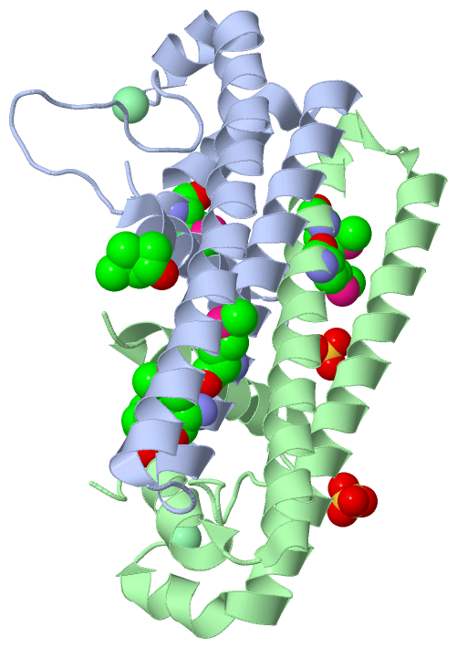
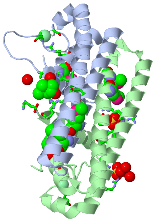
Chains, Units
Summary Information (see also Sequences/Alignments below) |
Ligands, Modified Residues, Ions (4, 11)| Asymmetric/Biological Unit (4, 11) |
Sites (5, 5)
Asymmetric Unit (5, 5)
|
SS Bonds (0, 0)| (no "SS Bond" information available for 2RDC) |
Cis Peptide Bonds (0, 0)| (no "Cis Peptide Bond" information available for 2RDC) |
SAPs(SNPs)/Variants (0, 0)| (no "SAP(SNP)/Variant" information available for 2RDC) |
PROSITE Motifs (0, 0)| (no "PROSITE Motif" information available for 2RDC) |
Exons (0, 0)| (no "Exon" information available for 2RDC) |
Sequences/Alignments
Asymmetric/Biological UnitChain A from PDB Type:PROTEIN Length:133 aligned with Q74H32_GEOSL | Q74H32 from UniProtKB/TrEMBL Length:152 Alignment length:133 29 39 49 59 69 79 89 99 109 119 129 139 149 Q74H32_GEOSL 20 ETTMGRYKKVIEITGHDEVAAKLLEGLIDAGTRYFSKVVEMEHRMASARFRLDGEELRELTETLDRSRRLAHESLISSLHVFNRYIVKEYGEELKEAGIEGGIFPKPEANRDRIAIADWAGELLTGIYENRHR 152 SCOP domains ------------------------------------------------------------------------------------------------------------------------------------- SCOP domains CATH domains 2rdcA00 A:20-152 protein ne1242 CATH domains Pfam domains ------------------------------------------------------------------------------------------------------------------------------------- Pfam domains SAPs(SNPs) ------------------------------------------------------------------------------------------------------------------------------------- SAPs(SNPs) PROSITE ------------------------------------------------------------------------------------------------------------------------------------- PROSITE Transcript ------------------------------------------------------------------------------------------------------------------------------------- Transcript 2rdc A 20 ETTmGRYKKVIEITGHDEVAAKLLEGLIDAGTRYFSKVVEmEHRmASARFRLDGEELRELTETLDRSRRLAHESLISSLHVFNRYIVKEYGEELKEAGIEGGIFPKPEANRDRIAIADWAGELLTGIYENRHR 152 | 29 39 49 59| | 69 79 89 99 109 119 129 139 149 | 60-MSE 23-MSE 64-MSE Chain B from PDB Type:PROTEIN Length:135 aligned with Q74H32_GEOSL | Q74H32 from UniProtKB/TrEMBL Length:152 Alignment length:135 27 37 47 57 67 77 87 97 107 117 127 137 147 Q74H32_GEOSL 18 ATETTMGRYKKVIEITGHDEVAAKLLEGLIDAGTRYFSKVVEMEHRMASARFRLDGEELRELTETLDRSRRLAHESLISSLHVFNRYIVKEYGEELKEAGIEGGIFPKPEANRDRIAIADWAGELLTGIYENRHR 152 SCOP domains --------------------------------------------------------------------------------------------------------------------------------------- SCOP domains CATH domains 2rdcB00 B:18-152 protein ne1242 CATH domains Pfam domains (1) DUF3232-2rdcB01 B:18-152 Pfam domains (1) Pfam domains (2) DUF3232-2rdcB02 B:18-152 Pfam domains (2) SAPs(SNPs) --------------------------------------------------------------------------------------------------------------------------------------- SAPs(SNPs) PROSITE --------------------------------------------------------------------------------------------------------------------------------------- PROSITE Transcript --------------------------------------------------------------------------------------------------------------------------------------- Transcript 2rdc B 18 ATETTmGRYKKVIEITGHDEVAAKLLEGLIDAGTRYFSKVVEmEHRmASARFRLDGEELRELTETLDRSRRLAHESLISSLHVFNRYIVKEYGEELKEAGIEGGIFPKPEANRDRIAIADWAGELLTGIYENRHR 152 | 27 37 47 57 | | 67 77 87 97 107 117 127 137 147 23-MSE 60-MSE 64-MSE
|
||||||||||||||||||||
SCOP Domains (0, 0)| (no "SCOP Domain" information available for 2RDC) |
CATH Domains (1, 2)
Asymmetric/Biological Unit
 |
Pfam Domains (1, 2)
Asymmetric/Biological Unit
|
Gene Ontology (3, 3)|
Asymmetric/Biological Unit(hide GO term definitions) Chain A,B (Q74H32_GEOSL | Q74H32)
|
||||||||||||||||||||||||||||||||||||
Interactive Views
|
|||||||||||||||||||||||||||||||||||||||||||||||||||||||||||||||||||||||||||||||||||||||||||||||||||||||||||||||||||||||||||||||||||||||||||||||||||||||||||||||||||||||
Still Images
|
||||||||||||||||
Databases
|
||||||||||||||||||||||||||||||||||||||||||||||||||||||||||||||||||||||||||||||||||||||||||||||||||||||||||||||||||||||||||||||||||||||||||||||||||||||||||||||||
Analysis Tools
|
|||||||||||||||||||||||||||||||||||||||||||||||||||||||||||||
Entries Sharing at Least One Protein Chain (UniProt ID)
Related Entries Specified in the PDB File
|
|