Show PDB file:   
         Plain Text   HTML   (compressed file size)
QuickSearch:   
by PDB,NDB,UniProt,PROSITE Code or Search Term(s)  
(-)Asymmetric Unit
(-)Asym. Unit - sites
(-)Biological Unit 1
(-)Biol. Unit 1 - sites
collapse expand < >
Image Asymmetric Unit
Asymmetric Unit  (Jmol Viewer)
Image Asym. Unit - sites
Asym. Unit - sites  (Jmol Viewer)
Image Biological Unit 1
Biological Unit 1  (Jmol Viewer)
Image Biol. Unit 1 - sites
Biol. Unit 1 - sites  (Jmol Viewer)

(-) Description

Title :  CRYSTAL STRUCTURE OF A PUTATIVE FLAVIN REDUCTASE (YCDH, HS_1225) FROM HAEMOPHILUS SOMNUS 129PT AT 1.06 A RESOLUTION
 
Authors :  Joint Center For Structural Genomics (Jcsg)
Date :  21 Aug 07  (Deposition) - 04 Sep 07  (Release) - 23 Mar 11  (Revision)
Method :  X-RAY DIFFRACTION
Resolution :  1.06
Chains :  Asym. Unit :  A
Biol. Unit 1:  A  (2x)
Keywords :  Split Barrel-Like, Structural Genomics, Joint Center For Structural Genomics, Jcsg, Protein Structure Initiative, Psi-2, Oxidoreductase (Keyword Search: [Gene Ontology, PubMed, Web (Google))
 
Reference :  Joint Center For Structural Genomics (Jcsg)
Crystal Structure Of Putative Flavin Reductase (Yp_719437. 1 From Haemophilus Somnus 129Pt At 1. 06 A Resolution
To Be Published
PubMed: search

(-) Compounds

Molecule 1 - POSSIBLE FLAVIN REDUCTASE
    ChainsA
    EngineeredYES
    Expression SystemESCHERICHIA COLI
    Expression System PlasmidSPEEDET
    Expression System StrainHK100
    Expression System Taxid562
    Expression System Vector TypePLASMID
    GeneYP_719437.1, YCDH
    Organism ScientificHAEMOPHILUS SOMNUS 129PT
    Organism Taxid205914
    Strain129PT

 Structural Features

(-) Chains, Units

  1
Asymmetric Unit A
Biological Unit 1 (2x)A

Summary Information (see also Sequences/Alignments below)

(-) Ligands, Modified Residues, Ions  (4, 14)

Asymmetric Unit (4, 14)
No.NameCountTypeFull Name
1ACT5Ligand/IonACETATE ION
2EDO3Ligand/Ion1,2-ETHANEDIOL
3MSE3Mod. Amino AcidSELENOMETHIONINE
4SO43Ligand/IonSULFATE ION
Biological Unit 1 (4, 28)
No.NameCountTypeFull Name
1ACT10Ligand/IonACETATE ION
2EDO6Ligand/Ion1,2-ETHANEDIOL
3MSE6Mod. Amino AcidSELENOMETHIONINE
4SO46Ligand/IonSULFATE ION

(-) Sites  (11, 11)

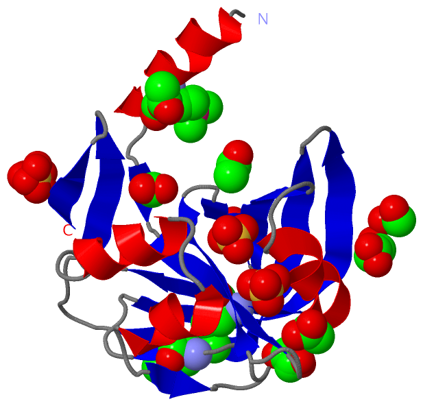
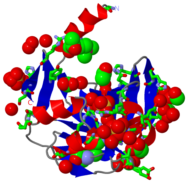
Asymmetric Unit (11, 11)
No.NameEvidenceResiduesDescription
01AC1SOFTWAREASN A:52 , HIS A:128 , HOH A:169BINDING SITE FOR RESIDUE ACT A 158
02AC2SOFTWARETYR A:149 , HOH A:301BINDING SITE FOR RESIDUE ACT A 159
03AC3SOFTWAREGLU A:59 , ARG A:152 , HOH A:221BINDING SITE FOR RESIDUE ACT A 160
04AC4SOFTWARESER A:39 , VAL A:40 , THR A:41 , ASP A:42 , HIS A:128 , HOH A:207 , HOH A:210BINDING SITE FOR RESIDUE ACT A 161
05AC5SOFTWAREGLY A:25 , GLU A:26 , THR A:27 , HIS A:30 , HIS A:60 , ASN A:64BINDING SITE FOR RESIDUE ACT A 162
06AC6SOFTWARELYS A:123 , HIS A:124 , HIS A:155 , SER A:156 , HOH A:194 , HOH A:233 , HOH A:289 , HOH A:303BINDING SITE FOR RESIDUE SO4 A 163
07AC7SOFTWAREGLY A:31 , PHE A:32 , ALA A:55 , ARG A:56 , ALA A:57 , ARG A:92 , HOH A:232 , HOH A:261BINDING SITE FOR RESIDUE SO4 A 164
08AC8SOFTWAREHIS A:30 , ARG A:56 , HIS A:60 , GLN A:89 , HOH A:213 , HOH A:223 , HOH A:231 , HOH A:264 , HOH A:288 , HOH A:290BINDING SITE FOR RESIDUE SO4 A 165
09AC9SOFTWAREALA A:74 , HIS A:124 , HIS A:155 , EDO A:167 , HOH A:194BINDING SITE FOR RESIDUE EDO A 166
10BC1SOFTWAREVAL A:62 , ARG A:65 , ALA A:74 , GLU A:75 , EDO A:166 , HOH A:183 , HOH A:222 , HOH A:245 , HOH A:246BINDING SITE FOR RESIDUE EDO A 167
11BC2SOFTWAREVAL A:2 , SER A:94 , LYS A:134 , HOH A:195 , HOH A:208 , HOH A:253 , HOH A:260BINDING SITE FOR RESIDUE EDO A 168

(-) SS Bonds  (1, 1)

Asymmetric Unit
No.Residues
1A:38 -A:38

(-) Cis Peptide Bonds  (1, 1)

Asymmetric Unit
No.Residues
1Ser A:43 -Pro A:44

 Sequence-Structure Mapping

(-) SAPs(SNPs)/Variants  (0, 0)

(no "SAP(SNP)/Variant" information available for 2R0X)

(-) PROSITE Motifs  (0, 0)

(no "PROSITE Motif" information available for 2R0X)

(-) Exons   (0, 0)

(no "Exon" information available for 2R0X)

(-) Sequences/Alignments

Asymmetric Unit
   Reformat: Number of residues per line =  ('0' or empty: single-line sequence representation)
  Number of residues per labelling interval =   
  UniProt sequence: complete  aligned part    
   Show mapping: SCOP domains CATH domains Pfam domains Secondary structure (by author)
SAPs(SNPs) PROSITE motifs Exons
(details for a mapped element are shown in a popup box when the mouse pointer rests over it)
Chain A from PDB  Type:PROTEIN  Length:156
 aligned with Q0I3S1_HAES1 | Q0I3S1 from UniProtKB/TrEMBL  Length:147

    Alignment length:156
                                     1                                                                                                                                                  
                                     1        11        21        31        41        51        61        71        81        91       101       111       121       131       141      
         Q0I3S1_HAES1     - ---------MAQLASAVHIVTTSGETGQHGFTASAVCSVTDSPPTLLVCINSNARAYEHFVKNRVLMVNTLTAEQSSLSNIFASPLSQEERFSNASWTTLTTGSPMLQDALINFDCEITEIKHVGTHDILICKIVDIHQSNAKNALVYRNRVYHSV 147
               SCOP domains d2r0xa_ A: automated matches                                                                                                                                 SCOP domains
               CATH domains 2r0xA00 A:2-157 Electron Transport, Fmn-binding Protein; Chain A                                                                                             CATH domains
               Pfam domains ---------Flavin_Reduct-2r0xA01 A:11-157                                                                                                                      Pfam domains
         Sec.struct. author .hhhhhhhhhhh..ee.eeeeee....eeeeee..eeeee....eeeeeee....hhhhhhhhheeeeee....hhhhhhhhh...hhhhhhh...ee.......ee....eeeeeeeeeeeee..eeeeeeeeeeeee.....eeeee..eeeee Sec.struct. author
                 SAPs(SNPs) ------------------------------------------------------------------------------------------------------------------------------------------------------------ SAPs(SNPs)
                    PROSITE ------------------------------------------------------------------------------------------------------------------------------------------------------------ PROSITE
                 Transcript ------------------------------------------------------------------------------------------------------------------------------------------------------------ Transcript
                 2r0x A   2 VEVSQFKDAmAQLASAVHIVTTSGETGQHGFTASAVCSVTDSPPTLLVCINSNARAYEHFVKNRVLmVNTLTAEQSSLSNIFASPLSQEERFSNASWTTLTTGSPmLQDALINFDCEITEIKHVGTHDILICKIVDIHQSNAKNALVYRNRVYHSV 157
                                    11        21        31        41        51        61      | 71        81        91       101     | 111       121       131       141       151      
                                    11-MSE                                                   68-MSE                                107-MSE                                              

   Legend:   → Mismatch (orange background)
  - → Gap (green background, '-', border residues have a numbering label)
    → Modified Residue (blue background, lower-case, 'x' indicates undefined single-letter code, labelled with number + name)
  x → Chemical Group (purple background, 'x', labelled with number + name, e.g. ACE or NH2)
  extra numbering lines below/above indicate numbering irregularities and modified residue names etc., number ends below/above '|'

 Classification and Annotation

(-) SCOP Domains  (1, 1)

Asymmetric Unit

(-) CATH Domains  (1, 1)

Asymmetric Unit
(-)
Class: Mainly Beta (13760)

(-) Pfam Domains  (1, 1)

Asymmetric Unit

(-) Gene Ontology  (3, 3)

Asymmetric Unit(hide GO term definitions)
Chain A   (Q0I3S1_HAES1 | Q0I3S1)
molecular function
    GO:0010181    FMN binding    Interacting selectively and non-covalently with flavin mono nucleotide. Flavin mono nucleotide (FMN) is the coenzyme or the prosthetic group of various flavoprotein oxidoreductase enzymes.
    GO:0016491    oxidoreductase activity    Catalysis of an oxidation-reduction (redox) reaction, a reversible chemical reaction in which the oxidation state of an atom or atoms within a molecule is altered. One substrate acts as a hydrogen or electron donor and becomes oxidized, while the other acts as hydrogen or electron acceptor and becomes reduced.
biological process
    GO:0055114    oxidation-reduction process    A metabolic process that results in the removal or addition of one or more electrons to or from a substance, with or without the concomitant removal or addition of a proton or protons.

 Visualization

(-) Interactive Views

Asymmetric Unit
  Complete Structure
    Jena3D(integrated viewing of ligand, site, SAP, PROSITE, SCOP information)
    WebMol | AstexViewer[tm]@PDBe
(Java Applets, require no local installation except for Java; loading may be slow)
    STRAP
(Java WebStart application, automatic local installation, requires Java; full application with system access!)
    RasMol
(require local installation)
    Molscript (VRML)
(requires installation of a VRML viewer; select preferred view via VRML and generate a mono or stereo PDF format file)
 
  Ligands, Modified Residues, Ions
    ACT  [ RasMol | Jena3D ]  +environment [ RasMol | Jena3D ]
    EDO  [ RasMol | Jena3D ]  +environment [ RasMol | Jena3D ]
    MSE  [ RasMol | Jena3D ]  +environment [ RasMol | Jena3D ]
    SO4  [ RasMol | Jena3D ]  +environment [ RasMol | Jena3D ]
 
  Sites
    AC1  [ RasMol ]  +environment [ RasMol ]
    AC2  [ RasMol ]  +environment [ RasMol ]
    AC3  [ RasMol ]  +environment [ RasMol ]
    AC4  [ RasMol ]  +environment [ RasMol ]
    AC5  [ RasMol ]  +environment [ RasMol ]
    AC6  [ RasMol ]  +environment [ RasMol ]
    AC7  [ RasMol ]  +environment [ RasMol ]
    AC8  [ RasMol ]  +environment [ RasMol ]
    AC9  [ RasMol ]  +environment [ RasMol ]
    BC1  [ RasMol ]  +environment [ RasMol ]
    BC2  [ RasMol ]  +environment [ RasMol ]
 
  Cis Peptide Bonds
    Ser A:43 - Pro A:44   [ RasMol ]  
 
Biological Unit
  Complete Structure
    Biological Unit 1  [ Jena3D ]

(-) Still Images

Jmol
  protein: cartoon or spacefill or dots and stick; nucleic acid: cartoon and stick; ligands: spacefill; active site: stick
Molscript
  protein, nucleic acid: cartoon; ligands: spacefill; active site: ball and stick

 Databases and Analysis Tools

(-) Databases

Access by PDB/NDB ID
  2r0x
    Family and Domain InformationProDom | SYSTERS
    General Structural InformationGlycoscienceDB | MMDB | NDB | OCA | PDB | PDBe | PDBj | PDBsum | PDBWiki | PQS | PROTEOPEDIA
    Orientation in MembranesOPM
    Protein SurfaceSURFACE
    Secondary StructureDSSP (structure derived) | HSSP (homology derived)
    Structural GenomicsGeneCensus
    Structural NeighboursCE | VAST
    Structure ClassificationCATH | Dali | SCOP
    Validation and Original DataBMRB Data View | BMRB Restraints Grid | EDS | PROCHECK | RECOORD | WHAT_CHECK
 
Access by UniProt ID/Accession number
  Q0I3S1_HAES1 | Q0I3S1
    Comparative Protein Structure ModelsModBase
    Genomic InformationEnsembl
    Protein-protein InteractionDIP
    Sequence, Family and Domain InformationInterPro | Pfam | SMART | UniProtKB/TrEMBL
 
Access by Enzyme Classificator   (EC Number)
  (no 'Enzyme Classificator' available)
    General Enzyme InformationBRENDA | EC-PDB | Enzyme | IntEnz
    PathwayKEGG | MetaCyc
 
Access by Disease Identifier   (MIM ID)
  (no 'MIM ID' available)
    Disease InformationOMIM
 
Access by GenAge ID
  (no 'GenAge ID' available)
    Age Related InformationGenAge

(-) Analysis Tools

Access by PDB/NDB ID
    Domain InformationXDom
    Interatomic Contacts of Structural UnitsCSU
    Ligand-protein ContactsLPC
    Protein CavitiescastP
    Sequence and Secondary StructurePDBCartoon
    Structure AlignmentSTRAP(Java WebStart application, automatic local installation, requires Java; full application with system access!)
    Structure and Sequence BrowserSTING
 
Access by UniProt ID/Accession number
  Q0I3S1_HAES1 | Q0I3S1
    Protein Disorder PredictionDisEMBL | FoldIndex | GLOBPLOT (for more information see DisProt)

 Related Entries

(-) Entries Sharing at Least One Protein Chain (UniProt ID)

(no "Entries Sharing at Least One Protein Chain" available for 2R0X)

(-) Related Entries Specified in the PDB File

(no "Related Entries Specified in the PDB File" available for 2R0X)