|
|
|
|
 Description
|
|
Compounds
|
||||||||||||||||||||||||||||||||||||||||||||||||||||||||
Chains, Units
Summary Information (see also Sequences/Alignments below) |
Ligands, Modified Residues, Ions (0, 0)| (no "Ligand,Modified Residues,Ions" information available for 2R0J) |
Sites (0, 0)| (no "Site" information available for 2R0J) |
SS Bonds (0, 0)| (no "SS Bond" information available for 2R0J) |
Cis Peptide Bonds (1, 1)
Asymmetric/Biological Unit
|
||||||||
SAPs(SNPs)/Variants (0, 0)| (no "SAP(SNP)/Variant" information available for 2R0J) |
PROSITE Motifs (0, 0)| (no "PROSITE Motif" information available for 2R0J) |
Exons (0, 0)| (no "Exon" information available for 2R0J) |
Sequences/Alignments
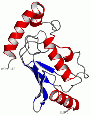
Asymmetric/Biological UnitChain A from PDB Type:PROTEIN Length:147 aligned with Q8I3J4_PLAF7 | Q8I3J4 from UniProtKB/TrEMBL Length:152 Alignment length:147 12 22 32 42 52 62 72 82 92 102 112 122 132 142 Q8I3J4_PLAF7 3 IPRRITKETQNLANEPPPGIMAVPVPENYRHFNILINGPDGTPYEGGTYKLELFLPEQYPMEPPKVRFLTKIYHPNIDKLGRICLDILKDKWSPALQIRTVLLSIQALLSSPEPDDPLDSKVAEHFKQDKNDAEHVARQWNKIYANN 149 SCOP domains d2r0ja_ A: automated matches SCOP domains CATH domains 2r0jA00 A:2-148 Ubiquitin Conjugating Enzyme CATH domains Pfam domains ---UQ_con-2r0jA01 A:5-142 ------ Pfam domains SAPs(SNPs) --------------------------------------------------------------------------------------------------------------------------------------------------- SAPs(SNPs) PROSITE --------------------------------------------------------------------------------------------------------------------------------------------------- PROSITE Transcript --------------------------------------------------------------------------------------------------------------------------------------------------- Transcript 2r0j A 2 IPRRITKETQNLANEPPPGIMAVPVPENYRHFNILINGPDGTPYEGGTYKLELFLPEQYPMEPPKVRFLTKIYHPNIDKLGRICLDILKDKWSPALQIRTVLLSIQALLSSPEPDDPLDSKVAEHFKQDKNDAEHVARQWNKIYANN 148 11 21 31 41 51 61 71 81 91 101 111 121 131 141
|
||||||||||||||||||||
SCOP Domains (1, 1)
Asymmetric/Biological Unit
 |
CATH Domains (1, 1)
Asymmetric/Biological Unit
|
Pfam Domains (1, 1)
Asymmetric/Biological Unit
|
Gene Ontology (9, 9)|
Asymmetric/Biological Unit(hide GO term definitions) Chain A (Q8I3J4_PLAF7 | Q8I3J4)
|
||||||||||||||||||||||||||||||||||||||||||||||||||||||||||||||||||||||||
Interactive Views
|
|||||||||||||||||||||||||||||||||||||||||||||||||||||||||||||||||||||||||||||||||||||||||||||||||||||||||||||||||||||
Still Images
|
||||||||||||||||
Databases
|
||||||||||||||||||||||||||||||||||||||||||||||||||||||||||||||||||||||||||||||||||||||||||||||||||||||||||||||||||||||||||||||||||||||||||||||||||||||||||||||||
Analysis Tools
|
|||||||||||||||||||||||||||||||||||||||||||||||||||||||||||||
Entries Sharing at Least One Protein Chain (UniProt ID)
Related Entries Specified in the PDB File
|
|