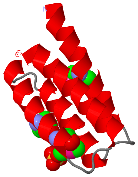
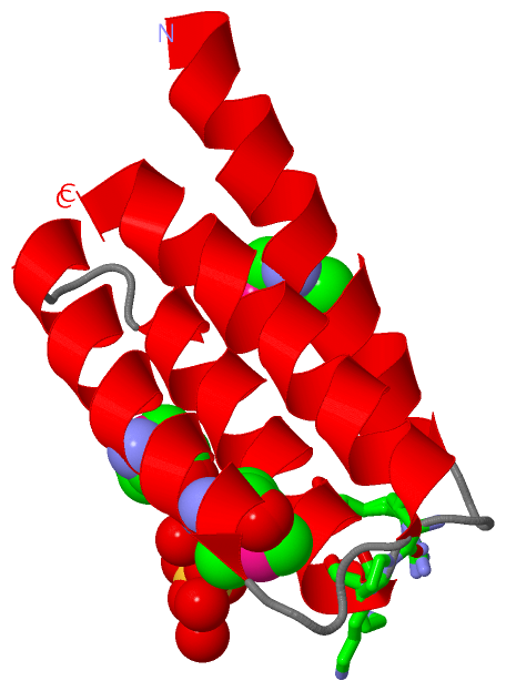
Show PDB file:   
         Plain Text   HTML   (compressed file size)
QuickSearch:   
by PDB,NDB,UniProt,PROSITE Code or Search Term(s)  
(-)Asym./Biol. Unit
(-)Asym./Biol. Unit - sites
collapse expand < >
Image Asym./Biol. Unit
Asym./Biol. Unit  (Jmol Viewer)
Image Asym./Biol. Unit - sites
Asym./Biol. Unit - sites  (Jmol Viewer)

(-) Description

Title :  CRYSTAL STRUCTURE OF A PROTEIN FROM UNCHARACTERIZED FAMILY UPF0147 FROM THERMOPLASMA ACIDOPHILUM
 
Authors :  M. E. Cuff, E. Duggan, M. Gu, A. Joachimiak, Midwest Center For Structural Genomics (Mcsg)
Date :  30 Jul 07  (Deposition) - 11 Sep 07  (Release) - 24 Feb 09  (Revision)
Method :  X-RAY DIFFRACTION
Resolution :  1.30
Chains :  Asym./Biol. Unit :  A
Keywords :  Structural Genomics, Four-Helix Bundle, Thermoplasma Acidophilum, Psi-2, Protein Structure Initiative, Midwest Center For Structural Genomics, Mcsg, Unknown Function (Keyword Search: [Gene Ontology, PubMed, Web (Google))
 
Reference :  M. E. Cuff, E. Duggan, M. Gu, A. Joachimiak
Structure Of A Protein From Uncharacterized Family Upf0147 From Thermoplasma Acidophilum.
To Be Published
PubMed: search
(for further references see the PDB file header)

(-) Compounds

Molecule 1 - UPF0147 PROTEIN TA0600
    Atcc25905
    ChainsA
    EngineeredYES
    Expression SystemESCHERICHIA COLI BL21(DE3)
    Expression System PlasmidPMCSG7
    Expression System StrainBL21(DE3)
    Expression System Taxid469008
    Expression System Vector TypePLASMID
    GeneCAC11739, TA0600
    Organism ScientificTHERMOPLASMA ACIDOPHILUM DSM 1728
    Organism Taxid273075
    StrainDSM 1728, IFO 15155, JCM 9062, AMRC-C165

 Structural Features

(-) Chains, Units

  1
Asymmetric/Biological Unit A

Summary Information (see also Sequences/Alignments below)

(-) Ligands, Modified Residues, Ions  (2, 4)

Asymmetric/Biological Unit (2, 4)
No.NameCountTypeFull Name
1MSE3Mod. Amino AcidSELENOMETHIONINE
2SO41Ligand/IonSULFATE ION

(-) Sites  (1, 1)

Asymmetric Unit (1, 1)
No.NameEvidenceResiduesDescription
1AC1SOFTWAREVAL A:25 , PRO A:26 , LYS A:27 , ARG A:30 , LYS A:31 , HOH A:106 , HOH A:112 , HOH A:212 , HOH A:215BINDING SITE FOR RESIDUE SO4 A 87

(-) SS Bonds  (0, 0)

(no "SS Bond" information available for 2QSB)

(-) Cis Peptide Bonds  (0, 0)

(no "Cis Peptide Bond" information available for 2QSB)

 Sequence-Structure Mapping

(-) SAPs(SNPs)/Variants  (0, 0)

(no "SAP(SNP)/Variant" information available for 2QSB)

(-) PROSITE Motifs  (0, 0)

(no "PROSITE Motif" information available for 2QSB)

(-) Exons   (0, 0)

(no "Exon" information available for 2QSB)

(-) Sequences/Alignments

Asymmetric/Biological Unit
   Reformat: Number of residues per line =  ('0' or empty: single-line sequence representation)
  Number of residues per labelling interval =   
  UniProt sequence: complete  aligned part    
   Show mapping: SCOP domains CATH domains Pfam domains Secondary structure (by author)
SAPs(SNPs) PROSITE motifs Exons
(details for a mapped element are shown in a popup box when the mouse pointer rests over it)
Chain A from PDB  Type:PROTEIN  Length:85
 aligned with Y600_THEAC | Q9HKJ8 from UniProtKB/Swiss-Prot  Length:88

    Alignment length:85
                                    11        21        31        41        51        61        71        81     
            Y600_THEAC    2 VRVDQNLFNEVMYLLDELSQDITVPKNVRKVAQDSKAKLSQENESLDLRCATVLSMLDEMANDPNVPAHGRTDLYTIISKLEALS 86
               SCOP domains d2qsba1 A:2-86 Uncharacterized protein Ta0600                                         SCOP domains
               CATH domains 2qsbA00 A:2-86 Ta0600-like domain                                                     CATH domains
               Pfam domains UPF0147-2qsbA01 A:2-86                                                                Pfam domains
         Sec.struct. author hhhhhhhhhhhhhhhhhhhh....hhhhhhhhhhhhhhhh....hhhhhhhhhhhhhhhhhh....hhhhhhhhhhhhhhhhhhh Sec.struct. author
                 SAPs(SNPs) ------------------------------------------------------------------------------------- SAPs(SNPs)
                    PROSITE ------------------------------------------------------------------------------------- PROSITE
                 Transcript ------------------------------------------------------------------------------------- Transcript
                  2qsb A  2 VRVDQNLFNEVmYLLDELSQDITVPKNVRKVAQDSKAKLSQENESLDLRCATVLSmLDEmANDPNVPAHGRTDLYTIISKLEALS 86
                                    11 |      21        31        41        51     |  61        71        81     
                                      13-MSE                                      57-MSE                         
                                                                                      61-MSE                     

   Legend:   → Mismatch (orange background)
  - → Gap (green background, '-', border residues have a numbering label)
    → Modified Residue (blue background, lower-case, 'x' indicates undefined single-letter code, labelled with number + name)
  x → Chemical Group (purple background, 'x', labelled with number + name, e.g. ACE or NH2)
  extra numbering lines below/above indicate numbering irregularities and modified residue names etc., number ends below/above '|'

 Classification and Annotation

(-) SCOP Domains  (1, 1)

Asymmetric/Biological Unit

(-) CATH Domains  (1, 1)

Asymmetric/Biological Unit

(-) Pfam Domains  (1, 1)

Asymmetric/Biological Unit

(-) Gene Ontology  (0, 0)

Asymmetric/Biological Unit(hide GO term definitions)
    (no "Gene Ontology" information available for 2QSB)

 Visualization

(-) Interactive Views

Asymmetric/Biological Unit
  Complete Structure
    Jena3D(integrated viewing of ligand, site, SAP, PROSITE, SCOP information)
    WebMol | AstexViewer[tm]@PDBe
(Java Applets, require no local installation except for Java; loading may be slow)
    STRAP
(Java WebStart application, automatic local installation, requires Java; full application with system access!)
    RasMol
(require local installation)
    Molscript (VRML)
(requires installation of a VRML viewer; select preferred view via VRML and generate a mono or stereo PDF format file)
 
  Ligands, Modified Residues, Ions
    MSE  [ RasMol | Jena3D ]  +environment [ RasMol | Jena3D ]
    SO4  [ RasMol | Jena3D ]  +environment [ RasMol | Jena3D ]
 
  Sites
    AC1  [ RasMol ]  +environment [ RasMol ]
 
  Cis Peptide Bonds
(no "Cis Peptide Bonds" information available for 2qsb)
 

(-) Still Images

Jmol
  protein: cartoon or spacefill or dots and stick; nucleic acid: cartoon and stick; ligands: spacefill; active site: stick
Molscript
  protein, nucleic acid: cartoon; ligands: spacefill; active site: ball and stick

 Databases and Analysis Tools

(-) Databases

Access by PDB/NDB ID
  2qsb
    Family and Domain InformationProDom | SYSTERS
    General Structural InformationGlycoscienceDB | MMDB | NDB | OCA | PDB | PDBe | PDBj | PDBsum | PDBWiki | PQS | PROTEOPEDIA
    Orientation in MembranesOPM
    Protein SurfaceSURFACE
    Secondary StructureDSSP (structure derived) | HSSP (homology derived)
    Structural GenomicsGeneCensus
    Structural NeighboursCE | VAST
    Structure ClassificationCATH | Dali | SCOP
    Validation and Original DataBMRB Data View | BMRB Restraints Grid | EDS | PROCHECK | RECOORD | WHAT_CHECK
 
Access by UniProt ID/Accession number
  Y600_THEAC | Q9HKJ8
    Comparative Protein Structure ModelsModBase
    Genomic InformationEnsembl
    Protein-protein InteractionDIP
    Sequence, Family and Domain InformationInterPro | Pfam | SMART | UniProtKB/SwissProt
 
Access by Enzyme Classificator   (EC Number)
  (no 'Enzyme Classificator' available)
    General Enzyme InformationBRENDA | EC-PDB | Enzyme | IntEnz
    PathwayKEGG | MetaCyc
 
Access by Disease Identifier   (MIM ID)
  (no 'MIM ID' available)
    Disease InformationOMIM
 
Access by GenAge ID
  (no 'GenAge ID' available)
    Age Related InformationGenAge

(-) Analysis Tools

Access by PDB/NDB ID
    Domain InformationXDom
    Interatomic Contacts of Structural UnitsCSU
    Ligand-protein ContactsLPC
    Protein CavitiescastP
    Sequence and Secondary StructurePDBCartoon
    Structure AlignmentSTRAP(Java WebStart application, automatic local installation, requires Java; full application with system access!)
    Structure and Sequence BrowserSTING
 
Access by UniProt ID/Accession number
  Y600_THEAC | Q9HKJ8
    Protein Disorder PredictionDisEMBL | FoldIndex | GLOBPLOT (for more information see DisProt)

 Related Entries

(-) Entries Sharing at Least One Protein Chain (UniProt ID)

(no "Entries Sharing at Least One Protein Chain" available for 2QSB)

(-) Related Entries Specified in the PDB File

(no "Related Entries Specified in the PDB File" available for 2QSB)