|
|
|
|
 Description
|
|
Compounds
|
||||||||||||||||||||||||||||||||||||||||||||||||
Chains, Units
Summary Information (see also Sequences/Alignments below) |
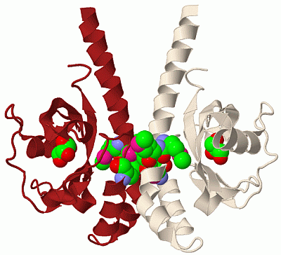
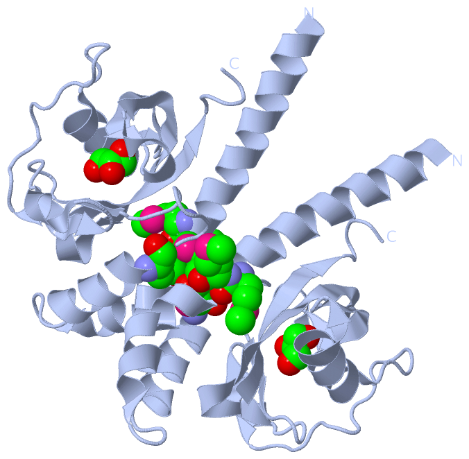
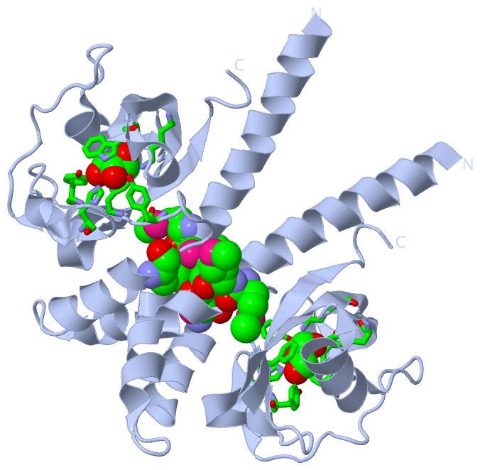
Ligands, Modified Residues, Ions (2, 5)| Asymmetric Unit (2, 5) Biological Unit 1 (2, 10) |
Sites (1, 1)
Asymmetric Unit (1, 1)
|
SS Bonds (0, 0)| (no "SS Bond" information available for 2QHK) |
Cis Peptide Bonds (0, 0)| (no "Cis Peptide Bond" information available for 2QHK) |
SAPs(SNPs)/Variants (0, 0)| (no "SAP(SNP)/Variant" information available for 2QHK) |
PROSITE Motifs (0, 0)| (no "PROSITE Motif" information available for 2QHK) |
Exons (0, 0)| (no "Exon" information available for 2QHK) |
Sequences/Alignments
Asymmetric UnitChain A from PDB Type:PROTEIN Length:148 aligned with Q87T87_VIBPA | Q87T87 from UniProtKB/TrEMBL Length:557 Alignment length:148 47 57 67 77 87 97 107 117 127 137 147 157 167 177 Q87T87_VIBPA 38 AELVRDRQELIDARKKELKAYMMMGVTAIKPLYDSDVNGSNKQAAKEILKAMRFESDGYFFAYDSQGINTLHAIKPSLEGKNLYDLKDENGVAVIAGLIDASQKGDGFLYFSWHKPTINAQAPKLGYAEYLQKWDWVLGTGIYIDDID 185 SCOP domains ---------------------------------------------------------------------------------------------------------------------------------------------------- SCOP domains CATH domains 2qhkA00 A:8-155 [code=3.30.450.20, no name defined] CATH domains Pfam domains ------Cache_2-2qhkA01 A:14-103 ---------------------------------------------------- Pfam domains SAPs(SNPs) ---------------------------------------------------------------------------------------------------------------------------------------------------- SAPs(SNPs) PROSITE ---------------------------------------------------------------------------------------------------------------------------------------------------- PROSITE Transcript ---------------------------------------------------------------------------------------------------------------------------------------------------- Transcript 2qhk A 8 AELVRDRQELIDARKKELKAYmmmGVTAIKPLYDSDVNGSNKQAAKEILKAmRFESDGYFFAYDSQGINTLHAIKPSLEGKNLYDLKDENGVAVIAGLIDASQKGDGFLYFSWHKPTINAQAPKLGYAEYLQKWDWVLGTGIYIDDID 155 17 27 ||| 37 47 57 | 67 77 87 97 107 117 127 137 147 29-MSE 59-MSE 30-MSE 31-MSE
|
||||||||||||||||||||
SCOP Domains (0, 0)| (no "SCOP Domain" information available for 2QHK) |
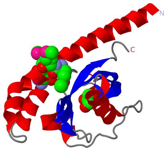
CATH Domains (1, 1)
Asymmetric Unit
 |
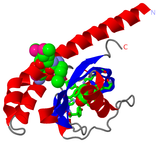
Pfam Domains (1, 1)
Asymmetric Unit
|
Gene Ontology (4, 4)|
Asymmetric Unit(hide GO term definitions) Chain A (Q87T87_VIBPA | Q87T87)
|
||||||||||||||||||||||||||||||||||||||||||
Interactive Views
|
|||||||||||||||||||||||||||||||||||||||||||||||||||||||||||||||||||||||||||||||||||||||||||||||||||||||||||||||||||||||||||||||||||||||||||||||
Still Images
|
||||||||||||||||
Databases
|
||||||||||||||||||||||||||||||||||||||||||||||||||||||||||||||||||||||||||||||||||||||||||||||||||||||||||||||||||||||||||||||||||||||||||||||||||||||||||||||||
Analysis Tools
|
|||||||||||||||||||||||||||||||||||||||||||||||||||||||||||||
Entries Sharing at Least One Protein Chain (UniProt ID)
Related Entries Specified in the PDB File
|
|