|
|
|
|
 Description
|
|
Compounds
|
||||||||||||||||||||||||||||||||||||||||||||
Chains, Units
Summary Information (see also Sequences/Alignments below) |
Ligands, Modified Residues, Ions (2, 2)| Asymmetric/Biological Unit (2, 2) |
Sites (2, 2)
Asymmetric Unit (2, 2)
|
SS Bonds (0, 0)| (no "SS Bond" information available for 2QDX) |
Cis Peptide Bonds (0, 0)| (no "Cis Peptide Bond" information available for 2QDX) |
SAPs(SNPs)/Variants (0, 0)| (no "SAP(SNP)/Variant" information available for 2QDX) |
PROSITE Motifs (0, 0)| (no "PROSITE Motif" information available for 2QDX) |
Exons (0, 0)| (no "Exon" information available for 2QDX) |
Sequences/Alignments
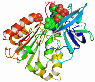
Asymmetric/Biological UnitChain A from PDB Type:PROTEIN Length:257 aligned with Q9HYK7_PSEAE | Q9HYK7 from UniProtKB/TrEMBL Length:258 Alignment length:257 11 21 31 41 51 61 71 81 91 101 111 121 131 141 151 161 171 181 191 201 211 221 231 241 251 Q9HYK7_PSEAE 2 SNLYTERVLSVHHWNDTLFSFKTTRNPGLRFKTGQFVMIGLEVDGRPLMRAYSIASPNYEEHLEFFSIKVPDGPLTSRLQHLKEGDELMVSRKPTGTLVHDDLLPGKHLYLLSTGTGMAPFLSVIQDPETYERYEKVILVHGVRWVSELAYADFITKVLPEHEYFGDQVKEKLIYYPLVTREPFRNQGRQTDLMRSGKLFEDIGLPPMNPQDDRAMICGSPSMLEETSAVLDSFGLKISPRMGEPGDYLIERAFVEK 258 SCOP domains d2qdxa1 A:2-100 Ferredoxin reductase (flavodoxin reductase) N-terminal domain d2qdxa2 A:101-258 Ferredoxin reductase (flavodoxin reductase) SCOP domains CATH domains 2qdxA01 A:2-96 Translation factors 2qdxA02 A:97-258 Nucleotide-binding domain of ferredoxin-NADP reductase (FNR) module CATH domains Pfam domains -------FAD_binding_6-2qdxA01 A:9-101 ----------NAD_binding_1-2qdxA02 A:112-230 ---------------------------- Pfam domains SAPs(SNPs) ----------------------------------------------------------------------------------------------------------------------------------------------------------------------------------------------------------------------------------------------------------------- SAPs(SNPs) PROSITE ----------------------------------------------------------------------------------------------------------------------------------------------------------------------------------------------------------------------------------------------------------------- PROSITE Transcript ----------------------------------------------------------------------------------------------------------------------------------------------------------------------------------------------------------------------------------------------------------------- Transcript 2qdx A 2 SNLYTERVLSVHHWNDTLFSFKTTRNPGLRFKTGQFVMIGLEVDGRPLMRAYSIASPNYEEHLEFFSIKVPDGPLTSRLQHLKEGDELMVSRKPTGTLVHDDLLPGKHLYLLSTGTGMAPFLSVIQDPETYERYEKVILVHGVRWVSELAYADFITKVLPEHEYFGDQVKEKLIYYPLVTREPFRNQGRQTDLMRSGKLFEDIGLPPMNPQDDRAMICGSPSMLEETSAVLDSFGLKISPRMGEPGDYLIERAFVEK 258 11 21 31 41 51 61 71 81 91 101 111 121 131 141 151 161 171 181 191 201 211 221 231 241 251
|
||||||||||||||||||||
SCOP Domains (2, 2)
Asymmetric/Biological Unit
 |
CATH Domains (2, 2)
Asymmetric/Biological Unit
|
Pfam Domains (2, 2)
Asymmetric/Biological Unit
|
Gene Ontology (3, 3)|
Asymmetric/Biological Unit(hide GO term definitions) Chain A (Q9HYK7_PSEAE | Q9HYK7)
|
||||||||||||||||||||||||||||||
Interactive Views
|
||||||||||||||||||||||||||||||||||||||||||||||||||||||||||||||||||||||||||||||||||||||||||||||||||||||||||||||||||||||||||||||||||||
Still Images
|
||||||||||||||||
Databases
|
||||||||||||||||||||||||||||||||||||||||||||||||||||||||||||||||||||||||||||||||||||||||||||||||||||||||||||||||||||||||||||||||||||||||||||||||||||||||||||||||
Analysis Tools
|
|||||||||||||||||||||||||||||||||||||||||||||||||||||||||||||
Entries Sharing at Least One Protein Chain (UniProt ID)
Related Entries Specified in the PDB File
|
|