Show PDB file:   
         Plain Text   HTML   (compressed file size)
QuickSearch:   
by PDB,NDB,UniProt,PROSITE Code or Search Term(s)  
(-)Biol.Unit 1 - manually
(-)Asym.Unit - manually
(-)Asymmetric Unit
(-)Asym. Unit - sites
(-)Biological Unit 1
(-)Biol. Unit 1 - sites
collapse expand < >
Image Biol.Unit 1 - manually
Biol.Unit 1 - manually  (Jmol Viewer)
Image Asym.Unit - manually
Asym.Unit - manually  (Jmol Viewer)
Image Asymmetric Unit
Asymmetric Unit  (Jmol Viewer)
Image Asym. Unit - sites
Asym. Unit - sites  (Jmol Viewer)
Image Biological Unit 1
Biological Unit 1  (Jmol Viewer)
Image Biol. Unit 1 - sites
Biol. Unit 1 - sites  (Jmol Viewer)

(-) Description

Title :  CRYSTAL STRUCTURE OF A DUF416 FAMILY PROTEIN (SBAL_3149) FROM SHEWANELLA BALTICA OS155 AT 1.91 A RESOLUTION
 
Authors :  Joint Center For Structural Genomics (Jcsg)
Date :  13 Jun 07  (Deposition) - 26 Jun 07  (Release) - 13 Jul 11  (Revision)
Method :  X-RAY DIFFRACTION
Resolution :  1.91
Chains :  Asym. Unit :  A
Biol. Unit 1:  A  (2x)
Keywords :  Structural Genomics, Joint Center For Structural Genomics, Jcsg, Protein Structure Initiative, Psi-2, Unknown Function (Keyword Search: [Gene Ontology, PubMed, Web (Google))
 
Reference :  Joint Center For Structural Genomics (Jcsg)
Crystal Structure Of Protein Of Unknown Function (Yp_001051499. 1) From Shewanella Baltica Os155 At 1. 91 A Resolution
To Be Published
PubMed: search

(-) Compounds

Molecule 1 - PROTEIN OF UNKNOWN FUNCTION
    ChainsA
    EngineeredYES
    Expression SystemESCHERICHIA COLI
    Expression System PlasmidSPEEDET
    Expression System StrainHK100
    Expression System Taxid562
    Expression System Vector TypePLASMID
    GeneYP_001051499.1, SBAL_3149
    Organism ScientificSHEWANELLA BALTICA
    Organism Taxid325240
    StrainOS155

 Structural Features

(-) Chains, Units

  1
Asymmetric Unit A
Biological Unit 1 (2x)A

Summary Information (see also Sequences/Alignments below)

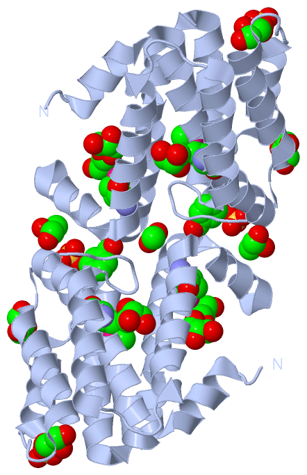
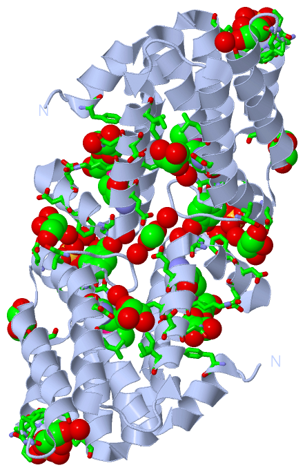
(-) Ligands, Modified Residues, Ions  (5, 13)

Asymmetric Unit (5, 13)
No.NameCountTypeFull Name
1ACT1Ligand/IonACETATE ION
2BEZ1Ligand/IonBENZOIC ACID
3GOL6Ligand/IonGLYCEROL
4MSE3Mod. Amino AcidSELENOMETHIONINE
5SO42Ligand/IonSULFATE ION
Biological Unit 1 (5, 26)
No.NameCountTypeFull Name
1ACT2Ligand/IonACETATE ION
2BEZ2Ligand/IonBENZOIC ACID
3GOL12Ligand/IonGLYCEROL
4MSE6Mod. Amino AcidSELENOMETHIONINE
5SO44Ligand/IonSULFATE ION

(-) Sites  (10, 10)

Asymmetric Unit (10, 10)
No.NameEvidenceResiduesDescription
01AC1SOFTWAREPHE A:86 , GLU A:87 , ALA A:88 , HOH A:274 , HOH A:297BINDING SITE FOR RESIDUE SO4 A 200
02AC2SOFTWAREPHE A:67 , ASN A:68 , ILE A:69 , ASP A:70 , GLN A:109 , HOH A:332BINDING SITE FOR RESIDUE SO4 A 201
03AC3SOFTWAREGLU A:156 , HOH A:271BINDING SITE FOR RESIDUE ACT A 202
04AC4SOFTWAREASN A:33 , GLY A:90 , SER A:135 , PHE A:147 , HIS A:151 , HOH A:213 , HOH A:215BINDING SITE FOR RESIDUE BEZ A 203
05AC5SOFTWARESER A:59 , LEU A:65 , LYS A:66 , PHE A:67 , HOH A:281BINDING SITE FOR RESIDUE GOL A 204
06AC6SOFTWAREPHE A:7 , LYS A:158 , ASN A:162 , GOL A:209BINDING SITE FOR RESIDUE GOL A 205
07AC7SOFTWARELEU A:160 , SER A:163 , ILE A:167 , LEU A:182 , ASP A:185 , HOH A:270BINDING SITE FOR RESIDUE GOL A 206
08AC8SOFTWAREGLU A:39 , ASP A:146 , PHE A:147 , VAL A:198 , HOH A:243BINDING SITE FOR RESIDUE GOL A 207
09AC9SOFTWAREGLY A:44 , ASN A:79BINDING SITE FOR RESIDUE GOL A 208
10BC1SOFTWAREASP A:145 , GOL A:205BINDING SITE FOR RESIDUE GOL A 209

(-) SS Bonds  (0, 0)

(no "SS Bond" information available for 2Q9R)

(-) Cis Peptide Bonds  (0, 0)

(no "Cis Peptide Bond" information available for 2Q9R)

 Sequence-Structure Mapping

(-) SAPs(SNPs)/Variants  (0, 0)

(no "SAP(SNP)/Variant" information available for 2Q9R)

(-) PROSITE Motifs  (0, 0)

(no "PROSITE Motif" information available for 2Q9R)

(-) Exons   (0, 0)

(no "Exon" information available for 2Q9R)

(-) Sequences/Alignments

Asymmetric Unit
   Reformat: Number of residues per line =  ('0' or empty: single-line sequence representation)
  Number of residues per labelling interval =   
  UniProt sequence: complete  aligned part    
   Show mapping: SCOP domains CATH domains Pfam domains Secondary structure (by author)
SAPs(SNPs) PROSITE motifs Exons
(details for a mapped element are shown in a popup box when the mouse pointer rests over it)
Chain A from PDB  Type:PROTEIN  Length:196
 aligned with A3D7B7_SHEB5 | A3D7B7 from UniProtKB/TrEMBL  Length:199

    Alignment length:196
                                    13        23        33        43        53        63        73        83        93       103       113       123       133       143       153       163       173       183       193      
         A3D7B7_SHEB5     4 KTGFFKRLKALTLPQKQLFATALCQRMLPNYQLFSEVCEFGDPAVLSTALELLWQSLYDPKLKFNIDVHLQRLEDNTPEPADFEAYGVYPAMDAVVAISTLLGAIQGKIEEDIVNISKLSSSTVANYIEAISDVDLVDEALDDFVFAHEVMEEEKELQNSLLEIIEENPKITAELVKGLRKDIIETGVSNIGISVA 199
               SCOP domains ---------------------------------------------------------------------------------------------------------------------------------------------------------------------------------------------------- SCOP domains
               CATH domains 2q9rA01 A:4-198 YP_001051499.1 domain like                                                                                                                                                         - CATH domains
               Pfam domains -DUF416-2q9rA01 A:5-198                                                                                                                                                                            - Pfam domains
         Sec.struct. author ..hhhhhhhhhhhhhhhhhhhhhhhhhhhhhhhhhhhhh..hhhhhhhhhhhhhhhhhh.....hhhhhhhhhhhh..hhhhh...hhhhhhhhhhhhhhhhhhhhhhhhhhhhhhhhhhhhhhhhhhhhhhh...hhhhhhhhhhhhhhhhhhhhhhhhhhhhhhh....hhhhhhhhhhhhhhhh......... Sec.struct. author
                 SAPs(SNPs) ---------------------------------------------------------------------------------------------------------------------------------------------------------------------------------------------------- SAPs(SNPs)
                    PROSITE ---------------------------------------------------------------------------------------------------------------------------------------------------------------------------------------------------- PROSITE
                 Transcript ---------------------------------------------------------------------------------------------------------------------------------------------------------------------------------------------------- Transcript
                 2q9r A   4 KTGFFKRLKALTLPQKQLFATALCQRmLPNYQLFSEVCEFGDPAVLSTALELLWQSLYDPKLKFNIDVHLQRLEDNTPEPADFEAYGVYPAmDAVVAISTLLGAIQGKIEEDIVNISKLSSSTVANYIEAISDVDLVDEALDDFVFAHEVmEEEKELQNSLLEIIEENPKITAELVKGLRKDIIETGVSNIGISVA 199
                                    13        23      | 33        43        53        63        73        83        93 |     103       113       123       133       143       153|      163       173       183       193      
                                                     30-MSE                                                           95-MSE                                                    154-MSE                                         

   Legend:   → Mismatch (orange background)
  - → Gap (green background, '-', border residues have a numbering label)
    → Modified Residue (blue background, lower-case, 'x' indicates undefined single-letter code, labelled with number + name)
  x → Chemical Group (purple background, 'x', labelled with number + name, e.g. ACE or NH2)
  extra numbering lines below/above indicate numbering irregularities and modified residue names etc., number ends below/above '|'

 Classification and Annotation

(-) SCOP Domains  (0, 0)

(no "SCOP Domain" information available for 2Q9R)

(-) CATH Domains  (1, 1)

Asymmetric Unit

(-) Pfam Domains  (1, 1)

Asymmetric Unit

(-) Gene Ontology  (0, 0)

Asymmetric Unit(hide GO term definitions)
    (no "Gene Ontology" information available for 2Q9R)

 Visualization

(-) Interactive Views

Asymmetric Unit
  Complete Structure
    Jena3D(integrated viewing of ligand, site, SAP, PROSITE, SCOP information)
    WebMol | AstexViewer[tm]@PDBe
(Java Applets, require no local installation except for Java; loading may be slow)
    STRAP
(Java WebStart application, automatic local installation, requires Java; full application with system access!)
    RasMol
(require local installation)
    Molscript (VRML)
(requires installation of a VRML viewer; select preferred view via VRML and generate a mono or stereo PDF format file)
 
  Ligands, Modified Residues, Ions
    ACT  [ RasMol | Jena3D ]  +environment [ RasMol | Jena3D ]
    BEZ  [ RasMol | Jena3D ]  +environment [ RasMol | Jena3D ]
    GOL  [ RasMol | Jena3D ]  +environment [ RasMol | Jena3D ]
    MSE  [ RasMol | Jena3D ]  +environment [ RasMol | Jena3D ]
    SO4  [ RasMol | Jena3D ]  +environment [ RasMol | Jena3D ]
 
  Sites
    AC1  [ RasMol ]  +environment [ RasMol ]
    AC2  [ RasMol ]  +environment [ RasMol ]
    AC3  [ RasMol ]  +environment [ RasMol ]
    AC4  [ RasMol ]  +environment [ RasMol ]
    AC5  [ RasMol ]  +environment [ RasMol ]
    AC6  [ RasMol ]  +environment [ RasMol ]
    AC7  [ RasMol ]  +environment [ RasMol ]
    AC8  [ RasMol ]  +environment [ RasMol ]
    AC9  [ RasMol ]  +environment [ RasMol ]
    BC1  [ RasMol ]  +environment [ RasMol ]
 
  Cis Peptide Bonds
(no "Cis Peptide Bonds" information available for 2q9r)
 
Biological Unit
  Complete Structure
    Biological Unit 1  [ Jena3D ]

(-) Still Images

Jmol
  protein: cartoon or spacefill or dots and stick; nucleic acid: cartoon and stick; ligands: spacefill; active site: stick
Molscript
  protein, nucleic acid: cartoon; ligands: spacefill; active site: ball and stick

 Databases and Analysis Tools

(-) Databases

Access by PDB/NDB ID
  2q9r
    Family and Domain InformationProDom | SYSTERS
    General Structural InformationGlycoscienceDB | MMDB | NDB | OCA | PDB | PDBe | PDBj | PDBsum | PDBWiki | PQS | PROTEOPEDIA
    Orientation in MembranesOPM
    Protein SurfaceSURFACE
    Secondary StructureDSSP (structure derived) | HSSP (homology derived)
    Structural GenomicsGeneCensus
    Structural NeighboursCE | VAST
    Structure ClassificationCATH | Dali | SCOP
    Validation and Original DataBMRB Data View | BMRB Restraints Grid | EDS | PROCHECK | RECOORD | WHAT_CHECK
 
Access by UniProt ID/Accession number
  A3D7B7_SHEB5 | A3D7B7
    Comparative Protein Structure ModelsModBase
    Genomic InformationEnsembl
    Protein-protein InteractionDIP
    Sequence, Family and Domain InformationInterPro | Pfam | SMART | UniProtKB/TrEMBL
 
Access by Enzyme Classificator   (EC Number)
  (no 'Enzyme Classificator' available)
    General Enzyme InformationBRENDA | EC-PDB | Enzyme | IntEnz
    PathwayKEGG | MetaCyc
 
Access by Disease Identifier   (MIM ID)
  (no 'MIM ID' available)
    Disease InformationOMIM
 
Access by GenAge ID
  (no 'GenAge ID' available)
    Age Related InformationGenAge

(-) Analysis Tools

Access by PDB/NDB ID
    Domain InformationXDom
    Interatomic Contacts of Structural UnitsCSU
    Ligand-protein ContactsLPC
    Protein CavitiescastP
    Sequence and Secondary StructurePDBCartoon
    Structure AlignmentSTRAP(Java WebStart application, automatic local installation, requires Java; full application with system access!)
    Structure and Sequence BrowserSTING
 
Access by UniProt ID/Accession number
  A3D7B7_SHEB5 | A3D7B7
    Protein Disorder PredictionDisEMBL | FoldIndex | GLOBPLOT (for more information see DisProt)

 Related Entries

(-) Entries Sharing at Least One Protein Chain (UniProt ID)

(no "Entries Sharing at Least One Protein Chain" available for 2Q9R)

(-) Related Entries Specified in the PDB File

(no "Related Entries Specified in the PDB File" available for 2Q9R)