|
|
|
|
 Description
|
|
Compounds
|
||||||||||||||||||||||||||||||||||||||||||||
Chains, Units
Summary Information (see also Sequences/Alignments below) |
Ligands, Modified Residues, Ions (2, 13)| Asymmetric Unit (2, 13) Biological Unit 1 (2, 6) Biological Unit 2 (2, 7) Biological Unit 3 (2, 13) |
Sites (13, 13)
Asymmetric Unit (13, 13)
|
SS Bonds (0, 0)| (no "SS Bond" information available for 2Q7A) |
Cis Peptide Bonds (0, 0)| (no "Cis Peptide Bond" information available for 2Q7A) |
SAPs(SNPs)/Variants (0, 0)| (no "SAP(SNP)/Variant" information available for 2Q7A) |
PROSITE Motifs (0, 0)| (no "PROSITE Motif" information available for 2Q7A) |
Exons (0, 0)| (no "Exon" information available for 2Q7A) |
Sequences/Alignments
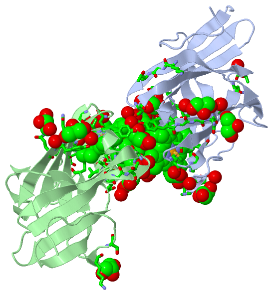
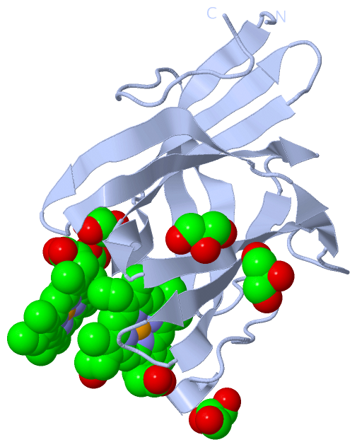
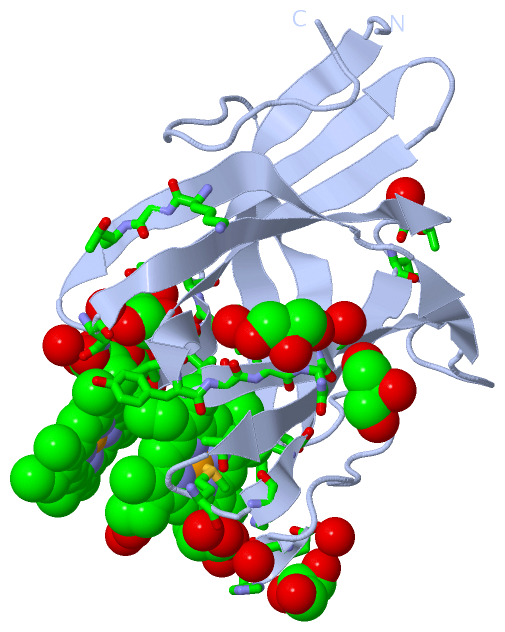
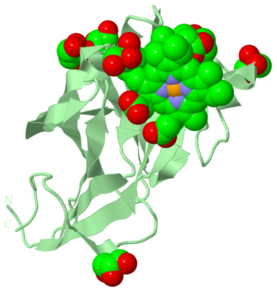
Asymmetric UnitChain A from PDB Type:PROTEIN Length:152 aligned with Q06A48_STRPY | Q06A48 from UniProtKB/TrEMBL Length:291 Alignment length:152 38 48 58 68 78 88 98 108 118 128 138 148 158 168 178 Q06A48_STRPY 29 ADKGQIYGCIIQRNYRHPISGQIEDSGGEHSFDIGQGMVEGTVYSDAMLEVSDAGKIVLTFRMSLADYSGNYQFWIQPGGTGSFQAVDYNITQKGTDTNGTTLDIAISLPTVNSIIRGSMFVEPMGREVVFYLSASELIQKYSGNMLAQLVT 180 SCOP domains -------------------------------------------------------------------------------------------------------------------------------------------------------- SCOP domains CATH domains -------------------------------------------------------------------------------------------------------------------------------------------------------- CATH domains Pfam domains -------------------------------------------------------------------------------------------------------------------------------------------------------- Pfam domains SAPs(SNPs) -------------------------------------------------------------------------------------------------------------------------------------------------------- SAPs(SNPs) PROSITE -------------------------------------------------------------------------------------------------------------------------------------------------------- PROSITE Transcript -------------------------------------------------------------------------------------------------------------------------------------------------------- Transcript 2q7a A 29 MDKGQIYGSIIQRNYRHPISGQIEDSGGEHSFDIGQGMVEGTVYSDAMLEVSDAGKIVLTFRMSLADYSGNYQFWIQPGGTGSFQAVDYNITQKGTDTNGTTLDIAISLPTVNSIIRGSMFVEPMGREVVFYLSASELIQKYSGNMLAQLVT 180 38 48 58 68 78 88 98 108 118 128 138 148 158 168 178 Chain B from PDB Type:PROTEIN Length:152 aligned with Q06A48_STRPY | Q06A48 from UniProtKB/TrEMBL Length:291 Alignment length:152 38 48 58 68 78 88 98 108 118 128 138 148 158 168 178 Q06A48_STRPY 29 ADKGQIYGCIIQRNYRHPISGQIEDSGGEHSFDIGQGMVEGTVYSDAMLEVSDAGKIVLTFRMSLADYSGNYQFWIQPGGTGSFQAVDYNITQKGTDTNGTTLDIAISLPTVNSIIRGSMFVEPMGREVVFYLSASELIQKYSGNMLAQLVT 180 SCOP domains -------------------------------------------------------------------------------------------------------------------------------------------------------- SCOP domains CATH domains -------------------------------------------------------------------------------------------------------------------------------------------------------- CATH domains Pfam domains (1) -HemeBinding_Shp-2q7aB01 B:30-180 Pfam domains (1) Pfam domains (2) -HemeBinding_Shp-2q7aB02 B:30-180 Pfam domains (2) SAPs(SNPs) -------------------------------------------------------------------------------------------------------------------------------------------------------- SAPs(SNPs) PROSITE -------------------------------------------------------------------------------------------------------------------------------------------------------- PROSITE Transcript -------------------------------------------------------------------------------------------------------------------------------------------------------- Transcript 2q7a B 29 MDKGQIYGSIIQRNYRHPISGQIEDSGGEHSFDIGQGMVEGTVYSDAMLEVSDAGKIVLTFRMSLADYSGNYQFWIQPGGTGSFQAVDYNITQKGTDTNGTTLDIAISLPTVNSIIRGSMFVEPMGREVVFYLSASELIQKYSGNMLAQLVT 180 38 48 58 68 78 88 98 108 118 128 138 148 158 168 178
|
||||||||||||||||||||
SCOP Domains (0, 0)| (no "SCOP Domain" information available for 2Q7A) |
CATH Domains (0, 0)| (no "CATH Domain" information available for 2Q7A) |
Pfam Domains (1, 2)
Asymmetric Unit

|
Gene Ontology (4, 4)|
Asymmetric Unit(hide GO term definitions) Chain A,B (Q06A48_STRPY | Q06A48)
|
||||||||||||||||||||||||||||||||||||
Interactive Views
|
|||||||||||||||||||||||||||||||||||||||||||||||||||||||||||||||||||||||||||||||||||||||||||||||||||||||||||||||||||||||||||||||||||||||||||||||||||||||||||||||||||||||||||||||||||||||||||||||||||||||||||||||||||||||||||||||||||||||||||||
Still Images
|
||||||||||||||||
Databases
|
||||||||||||||||||||||||||||||||||||||||||||||||||||||||||||||||||||||||||||||||||||||||||||||||||||||||||||||||||||||||||||||||||||||||||||||||||||||||||||||||
Analysis Tools
|
|||||||||||||||||||||||||||||||||||||||||||||||||||||||||||||
Entries Sharing at Least One Protein Chain (UniProt ID)
Related Entries Specified in the PDB File
|
|