Show PDB file:   
         Plain Text   HTML   (compressed file size)
QuickSearch:   
by PDB,NDB,UniProt,PROSITE Code or Search Term(s)  
(-)Asym./Biol. Unit
(-)Asym./Biol. Unit - sites
collapse expand < >
Image Asym./Biol. Unit
Asym./Biol. Unit  (Jmol Viewer)
Image Asym./Biol. Unit - sites
Asym./Biol. Unit - sites  (Jmol Viewer)

(-) Description

Title :  CRYSTAL STRUCTURES OF AN ISOLATED ABC-ATPASE IN COMPLEX WITH TNP-ADP
 
Authors :  C. Oswald, S. Jenewein, I. B. Holland, L. Schmitt
Date :  23 Apr 07  (Deposition) - 05 Feb 08  (Release) - 23 Sep 15  (Revision)
Method :  X-RAY DIFFRACTION
Resolution :  1.60
Chains :  Asym./Biol. Unit :  A
Keywords :  Abc-Transporter, Atpase, Nbd, Tnp-Nucleotide, Transport Protein (Keyword Search: [Gene Ontology, PubMed, Web (Google))
 
Reference :  C. Oswald, S. Jenewein, S. H. Smits, I. B. Holland, L. Schmitt
Water-Mediated Protein-Fluorophore Interactions Modulate Th Affinity Of An Abc-Atpase/Tnp-Adp Complex
J. Struct. Biol. V. 162 85 2008
PubMed-ID: 18155559  |  Reference-DOI: 10.1016/J.JSB.2007.11.006

(-) Compounds

Molecule 1 - ALPHA-HEMOLYSIN TRANSLOCATION ATP-BINDING PROTEIN HLYB
    ChainsA
    EngineeredYES
    Expression SystemESCHERICHIA COLI BL21(DE3)
    Expression System StrainBL21(DE3)
    Expression System Taxid469008
    FragmentABC TRANSPORTER (RESIDUES 467-707)
    GeneHLYB
    Organism ScientificESCHERICHIA COLI
    Organism Taxid562

 Structural Features

(-) Chains, Units

  1
Asymmetric/Biological Unit A

Summary Information (see also Sequences/Alignments below)

(-) Ligands, Modified Residues, Ions  (2, 2)

Asymmetric/Biological Unit (2, 2)
No.NameCountTypeFull Name
11281Ligand/IonSPIRO(2,4,6-TRINITROBENZENE[1,2A]-2O',3O'-METHYLENE-ADENINE-TRIPHOSPHATE
2ADP1Ligand/IonADENOSINE-5'-DIPHOSPHATE

(-) Sites  (2, 2)

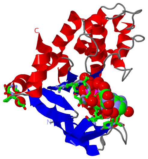
Asymmetric Unit (2, 2)
No.NameEvidenceResiduesDescription
1AC1SOFTWARETYR A:477 , PRO A:482 , ILE A:484 , GLY A:505 , SER A:506 , GLY A:507 , LYS A:508 , SER A:509 , THR A:510 , LYS A:513 , TYR A:519 , TRP A:540 , ARG A:543 , GLU A:689 , SER A:692 , GLU A:693 , PRO A:694 , HOH A:713 , HOH A:721 , HOH A:742 , HOH A:759 , HOH A:782 , HOH A:799 , HOH A:832 , HOH A:852BINDING SITE FOR RESIDUE 128 A 301
2AC2SOFTWARETYR A:477 , ILE A:484 , ARG A:503 , SER A:504 , GLY A:505 , SER A:506 , GLY A:507 , LYS A:508 , SER A:509 , THR A:510 , LYS A:513 , TYR A:519 , GLU A:689 , PRO A:694 , HOH A:713 , HOH A:721 , HOH A:742 , HOH A:759 , HOH A:782 , HOH A:801 , HOH A:832 , HOH A:852 , HOH A:898BINDING SITE FOR RESIDUE ADP A 401

(-) SS Bonds  (0, 0)

(no "SS Bond" information available for 2PMK)

(-) Cis Peptide Bonds  (0, 0)

(no "Cis Peptide Bond" information available for 2PMK)

 Sequence-Structure Mapping

(-) SAPs(SNPs)/Variants  (0, 0)

(no "SAP(SNP)/Variant" information available for 2PMK)

(-) PROSITE Motifs  (1, 1)

Asymmetric/Biological Unit (1, 1)
 PROSITEUniProtKBPDB
No.IDACDescriptionIDLocationCountLocation
1ABC_TRANSPORTER_1PS00211 ABC transporters family signature.HLYBP_ECOLX606-620  1A:606-620

(-) Exons   (0, 0)

(no "Exon" information available for 2PMK)

(-) Sequences/Alignments

Asymmetric/Biological Unit
   Reformat: Number of residues per line =  ('0' or empty: single-line sequence representation)
  Number of residues per labelling interval =   
  UniProt sequence: complete  aligned part    
   Show mapping: SCOP domains CATH domains Pfam domains Secondary structure (by author)
SAPs(SNPs) PROSITE motifs Exons
(details for a mapped element are shown in a popup box when the mouse pointer rests over it)
Chain A from PDB  Type:PROTEIN  Length:243
 aligned with HLYBP_ECOLX | P08716 from UniProtKB/Swiss-Prot  Length:707

    Alignment length:253
                                   464       474       484       494       504       514       524       534       544       554       564       574       584       594       604       614       624       634       644       654       664       674       684       694       704   
          HLYBP_ECOLX   455 YHGKLTLPEINGDITFRNIRFRYKPDSPVILDNINLSIKQGEVIGIVGRSGSGKSTLTKLIQRFYIPENGQVLIDGHDLALADPNWLRRQVGVVLQDNVLLNRSIIDNISLANPGMSVEKVIYAAKLAGAHDFISELREGYNTIVGEQGAGLSGGQRQRIAIARALVNNPKILIFDEATSALDYESEHVIMRNMHKICKGRTVIIIAHRLSTVKNADRIIVMEKGKIVEQGKHKELLSEPESLYSYLYQLQSD 707
               SCOP domains d2          pmka_ A: Haemolysin B ATP-binding protein                                                                                                                                                                                                         SCOP domains
               CATH domains ------------------------------------------------------------------------------------------------------------------------------------------------------------------------------------------------------------------------------------------------------------- CATH domains
               Pfam domains ------------------------------------------------------ABC_tran-2pmkA01 A:509-634                                                                                                    ------------------------------------------------------------------------- Pfam domains
         Sec.struct. author .e----------eeeeeeeeee......eeeeeeeeeee...eeeee.....hhhhhhhhhh......eeeeee..ee....hhhhhhhheeee.........hhhhhhh......hhhhhhhhhhhh.hhhhhh...hhhhh.........hhhhhhhhhhhhhhh....eeee.......hhhhhhhhhhhhhhhhh..eeeee..hhhhhh...eeeeee..eeeeeehhhhhhhh..hhhhhhhhhhh. Sec.struct. author
                 SAPs(SNPs) ------------------------------------------------------------------------------------------------------------------------------------------------------------------------------------------------------------------------------------------------------------- SAPs(SNPs)
                    PROSITE -------------------------------------------------------------------------------------------------------------------------------------------------------ABC_TRANSPORTER--------------------------------------------------------------------------------------- PROSITE
                 Transcript ------------------------------------------------------------------------------------------------------------------------------------------------------------------------------------------------------------------------------------------------------------- Transcript
                 2pmk A 465 HH----------DITFRNIRFRYKPDSPVILDNINLSIKQGEVIGIVGRSGSGKSTLTKLIQRFYIPENGQVLIDGHDLALADPNWLRRQVGVVLQDNVLLNRSIIDNISLANPGMSVEKVIYAAKLAGAHDFISELREGYNTIVGEQGAGLSGGQRQRIAIARALVNNPKILIFDEATSALDYESEHVIMRNMHKICKGRTVIIIAHRLSTVKNADRIIVMEKGKIVEQGKHKELLSEPESLYSYLYQLQSD 707
                             |       -  |    474       484       494       504       514       524       534       544       554       564       574       584       594       604       614       624       634       644       654       664       674       684       694       704   
                             |        467                                                                                                                                                                                                                                                
                           466                                                                                                                                                                                                                                                           

   Legend:   → Mismatch (orange background)
  - → Gap (green background, '-', border residues have a numbering label)
    → Modified Residue (blue background, lower-case, 'x' indicates undefined single-letter code, labelled with number + name)
  x → Chemical Group (purple background, 'x', labelled with number + name, e.g. ACE or NH2)
  extra numbering lines below/above indicate numbering irregularities and modified residue names etc., number ends below/above '|'

 Classification and Annotation

(-) SCOP Domains  (1, 1)

Asymmetric/Biological Unit

(-) CATH Domains  (0, 0)

(no "CATH Domain" information available for 2PMK)

(-) Pfam Domains  (1, 1)

Asymmetric/Biological Unit

(-) Gene Ontology  (15, 15)

Asymmetric/Biological Unit(hide GO term definitions)
Chain A   (HLYBP_ECOLX | P08716)
molecular function
    GO:0005524    ATP binding    Interacting selectively and non-covalently with ATP, adenosine 5'-triphosphate, a universally important coenzyme and enzyme regulator.
    GO:0016887    ATPase activity    Catalysis of the reaction: ATP + H2O = ADP + phosphate + 2 H+. May or may not be coupled to another reaction.
    GO:0042626    ATPase activity, coupled to transmembrane movement of substances    Catalysis of the reaction: ATP + H2O = ADP + phosphate, to directly drive the active transport of a substance across a membrane.
    GO:0016787    hydrolase activity    Catalysis of the hydrolysis of various bonds, e.g. C-O, C-N, C-C, phosphoric anhydride bonds, etc. Hydrolase is the systematic name for any enzyme of EC class 3.
    GO:0000166    nucleotide binding    Interacting selectively and non-covalently with a nucleotide, any compound consisting of a nucleoside that is esterified with (ortho)phosphate or an oligophosphate at any hydroxyl group on the ribose or deoxyribose.
    GO:0008233    peptidase activity    Catalysis of the hydrolysis of a peptide bond. A peptide bond is a covalent bond formed when the carbon atom from the carboxyl group of one amino acid shares electrons with the nitrogen atom from the amino group of a second amino acid.
    GO:0008565    protein transporter activity    Enables the directed movement of proteins into, out of or within a cell, or between cells.
biological process
    GO:0030253    protein secretion by the type I secretion system    The process in which proteins are secreted into the extracellular milieu via the type I secretion system; secretion occurs in a continuous process without the distinct presence of periplasmic intermediates and does not involve proteolytic processing of secreted proteins.
    GO:0006508    proteolysis    The hydrolysis of proteins into smaller polypeptides and/or amino acids by cleavage of their peptide bonds.
    GO:0055085    transmembrane transport    The process in which a solute is transported across a lipid bilayer, from one side of a membrane to the other
    GO:0006810    transport    The directed movement of substances (such as macromolecules, small molecules, ions) or cellular components (such as complexes and organelles) into, out of or within a cell, or between cells, or within a multicellular organism by means of some agent such as a transporter, pore or motor protein.
cellular component
    GO:0016021    integral component of membrane    The component of a membrane consisting of the gene products and protein complexes having at least some part of their peptide sequence embedded in the hydrophobic region of the membrane.
    GO:0016020    membrane    A lipid bilayer along with all the proteins and protein complexes embedded in it an attached to it.
    GO:0005886    plasma membrane    The membrane surrounding a cell that separates the cell from its external environment. It consists of a phospholipid bilayer and associated proteins.
    GO:0030256    type I protein secretion system complex    A complex of three secretory proteins that carry out secretion in the type I secretion system: an inner membrane transport ATPase (termed ABC protein for ATP-binding cassette), which provides the energy for protein secretion; an outer membrane protein, which is exported via the sec pathway; and a membrane fusion protein, which is anchored in the inner membrane and spans the periplasmic space.

 Visualization

(-) Interactive Views

Asymmetric/Biological Unit
  Complete Structure
    Jena3D(integrated viewing of ligand, site, SAP, PROSITE, SCOP information)
    WebMol | AstexViewer[tm]@PDBe
(Java Applets, require no local installation except for Java; loading may be slow)
    STRAP
(Java WebStart application, automatic local installation, requires Java; full application with system access!)
    RasMol
(require local installation)
    Molscript (VRML)
(requires installation of a VRML viewer; select preferred view via VRML and generate a mono or stereo PDF format file)
 
  Ligands, Modified Residues, Ions
    128  [ RasMol | Jena3D ]  +environment [ RasMol | Jena3D ]
    ADP  [ RasMol | Jena3D ]  +environment [ RasMol | Jena3D ]
 
  Sites
    AC1  [ RasMol ]  +environment [ RasMol ]
    AC2  [ RasMol ]  +environment [ RasMol ]
 
  Cis Peptide Bonds
(no "Cis Peptide Bonds" information available for 2pmk)
 

(-) Still Images

Jmol
  protein: cartoon or spacefill or dots and stick; nucleic acid: cartoon and stick; ligands: spacefill; active site: stick
Molscript
  protein, nucleic acid: cartoon; ligands: spacefill; active site: ball and stick

 Databases and Analysis Tools

(-) Databases

Access by PDB/NDB ID
  2pmk
    Family and Domain InformationProDom | SYSTERS
    General Structural InformationGlycoscienceDB | MMDB | NDB | OCA | PDB | PDBe | PDBj | PDBsum | PDBWiki | PQS | PROTEOPEDIA
    Orientation in MembranesOPM
    Protein SurfaceSURFACE
    Secondary StructureDSSP (structure derived) | HSSP (homology derived)
    Structural GenomicsGeneCensus
    Structural NeighboursCE | VAST
    Structure ClassificationCATH | Dali | SCOP
    Validation and Original DataBMRB Data View | BMRB Restraints Grid | EDS | PROCHECK | RECOORD | WHAT_CHECK
 
Access by UniProt ID/Accession number
  HLYBP_ECOLX | P08716
    Comparative Protein Structure ModelsModBase
    Genomic InformationEnsembl
    Protein-protein InteractionDIP
    Sequence, Family and Domain InformationInterPro | Pfam | SMART | UniProtKB/SwissProt
 
Access by Enzyme Classificator   (EC Number)
  (no 'Enzyme Classificator' available)
    General Enzyme InformationBRENDA | EC-PDB | Enzyme | IntEnz
    PathwayKEGG | MetaCyc
 
Access by Disease Identifier   (MIM ID)
  (no 'MIM ID' available)
    Disease InformationOMIM
 
Access by GenAge ID
  (no 'GenAge ID' available)
    Age Related InformationGenAge

(-) Analysis Tools

Access by PDB/NDB ID
    Domain InformationXDom
    Interatomic Contacts of Structural UnitsCSU
    Ligand-protein ContactsLPC
    Protein CavitiescastP
    Sequence and Secondary StructurePDBCartoon
    Structure AlignmentSTRAP(Java WebStart application, automatic local installation, requires Java; full application with system access!)
    Structure and Sequence BrowserSTING
 
Access by UniProt ID/Accession number
  HLYBP_ECOLX | P08716
    Protein Disorder PredictionDisEMBL | FoldIndex | GLOBPLOT (for more information see DisProt)

 Related Entries

(-) Entries Sharing at Least One Protein Chain (UniProt ID)

UniProtKB/Swiss-Prot
        HLYBP_ECOLX | P087161mt0 1xef 2ff7 2ffa 2ffb 2fgj 2fgk 3b5j

(-) Related Entries Specified in the PDB File

1mt0 1xef 2ff7