|
|
|
|
 Description
|
|
Compounds
|
||||||||||||||||||||||||||||||||||||||||||||||||
Chains, Units
Summary Information (see also Sequences/Alignments below) |
Ligands, Modified Residues, Ions (1, 1)
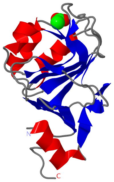
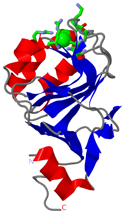
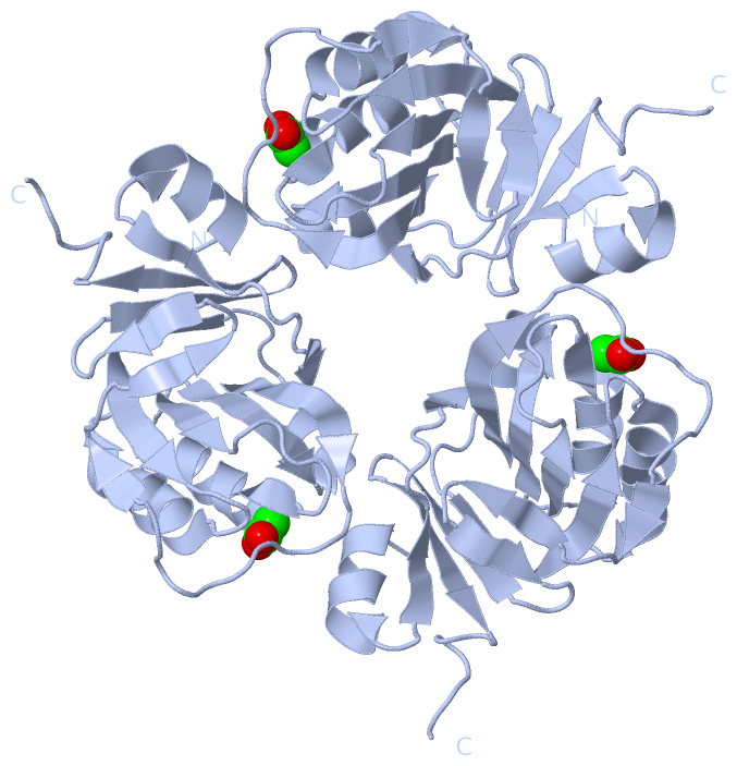
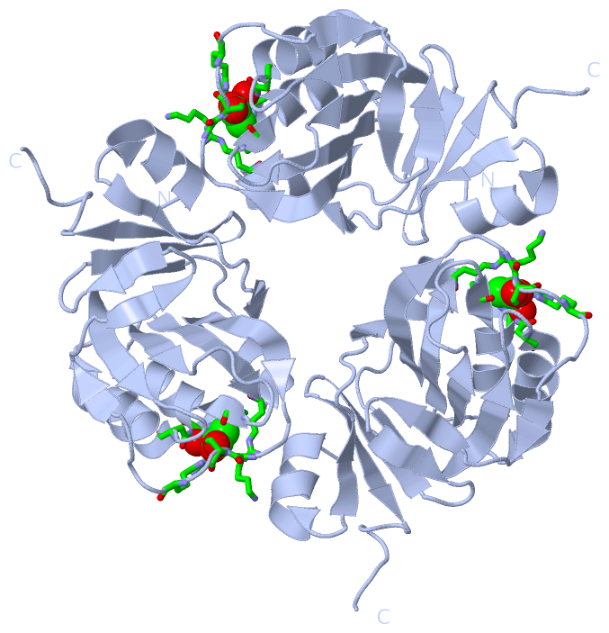
Asymmetric Unit (1, 1)
|
Sites (1, 1)
Asymmetric Unit (1, 1)
|
SS Bonds (0, 0)| (no "SS Bond" information available for 2PCN) |
Cis Peptide Bonds (0, 0)| (no "Cis Peptide Bond" information available for 2PCN) |
SAPs(SNPs)/Variants (0, 0)| (no "SAP(SNP)/Variant" information available for 2PCN) |
PROSITE Motifs (0, 0)| (no "PROSITE Motif" information available for 2PCN) |
Exons (0, 0)| (no "Exon" information available for 2PCN) |
Sequences/Alignments
Asymmetric UnitChain A from PDB Type:PROTEIN Length:161 aligned with Q5KYY8_GEOKA | Q5KYY8 from UniProtKB/TrEMBL Length:161 Alignment length:161 10 20 30 40 50 60 70 80 90 100 110 120 130 140 150 160 Q5KYY8_GEOKA 1 MKTADLCDQFLDELQVCELPFQSYGGKRMFSGPIATVDVFEDNVLVREALETVPPGTVLVVDGKGSRRVALLGDRLAQIACERGLAGVIIHGCIRDSAEIGAMPIGVMAIGTCPVKSKKEGKGARDVVLEFGGVRWEPGAYVYADADGVVVANKDLLAKNG 161 SCOP domains ----------------------------------------------------------------------------------------------------------------------------------------------------------------- SCOP domains CATH domains 2pcnA00 A:3-163 [code=3.50.30.40, no name defined] CATH domains Pfam domains Methyltransf_6-2pcnA01 A:3-153 ---------- Pfam domains SAPs(SNPs) ----------------------------------------------------------------------------------------------------------------------------------------------------------------- SAPs(SNPs) PROSITE ----------------------------------------------------------------------------------------------------------------------------------------------------------------- PROSITE Transcript ----------------------------------------------------------------------------------------------------------------------------------------------------------------- Transcript 2pcn A 3 MKTADLCDQFLDELQVCELPFQSYGGKRMFSGPIATVDVFEDNVLVREALETVPPGTVLVVDGKGSRRVALLGDRLAQIACERGLAGVIIHGCIRDSAEIGAMPIGVMAIGTCPVKSKKEGKGARDVVLEFGGVRWEPGAYVYADADGVVVANKDLLAKNG 163 12 22 32 42 52 62 72 82 92 102 112 122 132 142 152 162
|
||||||||||||||||||||
SCOP Domains (0, 0)| (no "SCOP Domain" information available for 2PCN) |
CATH Domains (1, 1)
Asymmetric Unit
 |
Pfam Domains (1, 1)
Asymmetric Unit
|
Gene Ontology (10, 10)|
Asymmetric Unit(hide GO term definitions) Chain A (Q5KYY8_GEOKA | Q5KYY8)
|
||||||||||||||||||||||||||||||||||||||||||||||||||||||||||||||||||||||||
Interactive Views
|
||||||||||||||||||||||||||||||||||||||||||||||||||||||||||||||||||||||||||||||||||||||||||||||||||||||||||||||||||||||||||||||||||||||||
Still Images
|
||||||||||||||||
Databases
|
||||||||||||||||||||||||||||||||||||||||||||||||||||||||||||||||||||||||||||||||||||||||||||||||||||||||||||||||||||||||||||||||||||||||||||||||||||||||||||||||
Analysis Tools
|
|||||||||||||||||||||||||||||||||||||||||||||||||||||||||||||
Entries Sharing at Least One Protein Chain (UniProt ID)
Related Entries Specified in the PDB File
|
|