Show PDB file:   
         Plain Text   HTML   (compressed file size)
QuickSearch:   
by PDB,NDB,UniProt,PROSITE Code or Search Term(s)  
(-)NMR Structure - model 1
(-)NMR Structure - all models
collapse expand < >
Image NMR Structure - model 1
NMR Structure - model 1  (Jmol Viewer)
Image NMR Structure - all models
NMR Structure - all models  (Jmol Viewer)

(-) Description

Title :  CALCIUM BINDING PROTEIN IN THE FREE FORM
 
Authors :  X. Zhang, Y. Hu, C. Jin
Date :  28 Feb 07  (Deposition) - 25 Mar 08  (Release) - 13 Jul 11  (Revision)
Method :  SOLUTION NMR
Resolution :  NOT APPLICABLE
Chains :  NMR Structure  :  A  (20x)
NMR Structure *:  A  (1x)
Keywords :  Calcium Binding, Metal Binding Protein (Keyword Search: [Gene Ontology, PubMed, Web (Google))
 
Reference :  X. Zhang, Y. Hu, B. Xia, C. Jin
Solution Structures Of Ccbp From Anabaena Reveals A New Fol And Novel Calcium Binding Sites
To Be Published
PubMed: search

(-) Compounds

Molecule 1 - ALR1010 PROTEIN
    ChainsA
    EngineeredYES
    Expression SystemESCHERICHIA COLI BL21(DE3)
    Expression System PlasmidPET15B
    Expression System StrainBL21(DE3)
    Expression System Taxid469008
    Expression System Vector TypePLASMID
    GeneCCBP
    Organism ScientificNOSTOC SP.
    Organism Taxid103690
    StrainPCC 7120
    SynonymCCBP

 Structural Features

(-) Chains, Units

  1
NMR Structure (20x)A
NMR Structure * (1x)A

Summary Information (see also Sequences/Alignments below)

(-) Ligands, Modified Residues, Ions  (0, 0)

(no "Ligand,Modified Residues,Ions" information available for 2P0P)

(-) Sites  (0, 0)

(no "Site" information available for 2P0P)

(-) SS Bonds  (0, 0)

(no "SS Bond" information available for 2P0P)

(-) Cis Peptide Bonds  (1, 20)
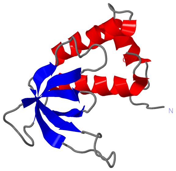
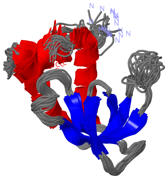
NMR Structure
No.ModelResidues
11, 2, 3, 4, 5, 6, 7, 8, 9, 10, 11, 12, 13, 14, 15, 16, 17, 18, 19, 20Phe A:42 -Pro A:43

 Sequence-Structure Mapping

(-) SAPs(SNPs)/Variants  (0, 0)

(no "SAP(SNP)/Variant" information available for 2P0P)

(-) PROSITE Motifs  (0, 0)

(no "PROSITE Motif" information available for 2P0P)

(-) Exons   (0, 0)

(no "Exon" information available for 2P0P)

(-) Sequences/Alignments

NMR Structure
   Reformat: Number of residues per line =  ('0' or empty: single-line sequence representation)
  Number of residues per labelling interval =   
  UniProt sequence: complete  aligned part    
   Show mapping: SCOP domains CATH domains Pfam domains Secondary structure (by author)
SAPs(SNPs) PROSITE motifs Exons
(details for a mapped element are shown in a popup box when the mouse pointer rests over it)
Chain A from PDB  Type:PROTEIN  Length:126
 aligned with Q8YY42_NOSS1 | Q8YY42 from UniProtKB/TrEMBL  Length:126

    Alignment length:126
                                    10        20        30        40        50        60        70        80        90       100       110       120      
         Q8YY42_NOSS1     1 MASVERDETREHRIETEIIVDAEDKEERAMGWYYYLDDTLEFPFMGKWKKKSRKTSTIEEKTVEVLGMAPDDECLKDMYVEVADIGGKDDDVYTAKLSDIEAIDVDDDTQEAIADWLYWLARGYKF 126
               SCOP domains ------------------------------------------------------------------------------------------------------------------------------ SCOP domains
               CATH domains ------------------------------------------------------------------------------------------------------------------------------ CATH domains
               Pfam domains -----------Calci_bind_CcbP-2p0pA01 A:12-121                                                                              ----- Pfam domains
         Sec.struct. author .......hhhhhhhhhhh.....hhhhhhhhhhhhhhhhh...eeeee............eeeeeeee..........eeeee........eeeee...eeee..hhhhhhhhhhhhhhhhhh... Sec.struct. author
                 SAPs(SNPs) ------------------------------------------------------------------------------------------------------------------------------ SAPs(SNPs)
                    PROSITE ------------------------------------------------------------------------------------------------------------------------------ PROSITE
                 Transcript ------------------------------------------------------------------------------------------------------------------------------ Transcript
                 2p0p A   1 MASVERDETREHRIETEIIVDAEDKEERAMGWYYYLDDTLEFPFMGKWKKKSRKTSTIEEKTVEVLGMAPDDECLKDMYVEVADIGGKDDDVYTAKLSDIEAIDVDDDTQEAIADWLYWLARGYKF 126
                                    10        20        30        40        50        60        70        80        90       100       110       120      

   Legend:   → Mismatch (orange background)
  - → Gap (green background, '-', border residues have a numbering label)
    → Modified Residue (blue background, lower-case, 'x' indicates undefined single-letter code, labelled with number + name)
  x → Chemical Group (purple background, 'x', labelled with number + name, e.g. ACE or NH2)
  extra numbering lines below/above indicate numbering irregularities and modified residue names etc., number ends below/above '|'

 Classification and Annotation

(-) SCOP Domains  (0, 0)

(no "SCOP Domain" information available for 2P0P)

(-) CATH Domains  (0, 0)

(no "CATH Domain" information available for 2P0P)

(-) Pfam Domains  (1, 1)

NMR Structure

(-) Gene Ontology  (1, 1)

NMR Structure(hide GO term definitions)
Chain A   (Q8YY42_NOSS1 | Q8YY42)
molecular function
    GO:0046872    metal ion binding    Interacting selectively and non-covalently with any metal ion.

 Visualization

(-) Interactive Views

NMR Structure
  Complete Structure
    Jena3D(integrated viewing of ligand, site, SAP, PROSITE, SCOP information)
    WebMol | AstexViewer[tm]@PDBe
(Java Applets, require no local installation except for Java; loading may be slow)
    STRAP
(Java WebStart application, automatic local installation, requires Java; full application with system access!)
    RasMol
(require local installation)
    Molscript (VRML)
(requires installation of a VRML viewer; select preferred view via VRML and generate a mono or stereo PDF format file)
 
  Ligands, Modified Residues, Ions
(no "Ligands, Modified Residues, Ions" information available for 2p0p)
 
  Sites
(no "Sites" information available for 2p0p)
 
  Cis Peptide Bonds
    Phe A:42 - Pro A:43   [ RasMol ]  
 

(-) Still Images

Jmol
  protein: cartoon or spacefill or dots and stick; nucleic acid: cartoon and stick; ligands: spacefill; active site: stick
Molscript
  protein, nucleic acid: cartoon; ligands: spacefill; active site: ball and stick

 Databases and Analysis Tools

(-) Databases

Access by PDB/NDB ID
  2p0p
    Family and Domain InformationProDom | SYSTERS
    General Structural InformationGlycoscienceDB | MMDB | NDB | OCA | PDB | PDBe | PDBj | PDBsum | PDBWiki | PQS | PROTEOPEDIA
    Orientation in MembranesOPM
    Protein SurfaceSURFACE
    Secondary StructureDSSP (structure derived) | HSSP (homology derived)
    Structural GenomicsGeneCensus
    Structural NeighboursCE | VAST
    Structure ClassificationCATH | Dali | SCOP
    Validation and Original DataBMRB Data View | BMRB Restraints Grid | EDS | PROCHECK | RECOORD | WHAT_CHECK
 
Access by UniProt ID/Accession number
  Q8YY42_NOSS1 | Q8YY42
    Comparative Protein Structure ModelsModBase
    Genomic InformationEnsembl
    Protein-protein InteractionDIP
    Sequence, Family and Domain InformationInterPro | Pfam | SMART | UniProtKB/TrEMBL
 
Access by Enzyme Classificator   (EC Number)
  (no 'Enzyme Classificator' available)
    General Enzyme InformationBRENDA | EC-PDB | Enzyme | IntEnz
    PathwayKEGG | MetaCyc
 
Access by Disease Identifier   (MIM ID)
  (no 'MIM ID' available)
    Disease InformationOMIM
 
Access by GenAge ID
  (no 'GenAge ID' available)
    Age Related InformationGenAge

(-) Analysis Tools

Access by PDB/NDB ID
    Domain InformationXDom
    Interatomic Contacts of Structural UnitsCSU
    Ligand-protein ContactsLPC
    Protein CavitiescastP
    Sequence and Secondary StructurePDBCartoon
    Structure AlignmentSTRAP(Java WebStart application, automatic local installation, requires Java; full application with system access!)
    Structure and Sequence BrowserSTING
 
Access by UniProt ID/Accession number
  Q8YY42_NOSS1 | Q8YY42
    Protein Disorder PredictionDisEMBL | FoldIndex | GLOBPLOT (for more information see DisProt)

 Related Entries

(-) Entries Sharing at Least One Protein Chain (UniProt ID)

UniProtKB/TrEMBL
        Q8YY42_NOSS1 | Q8YY422k2v 2p0q 3urg

(-) Related Entries Specified in the PDB File

2p0q