|
|
|
|
 Description
|
|
Compounds
|
||||||||||||||||||||||||||||||||||||||||||||||||||||
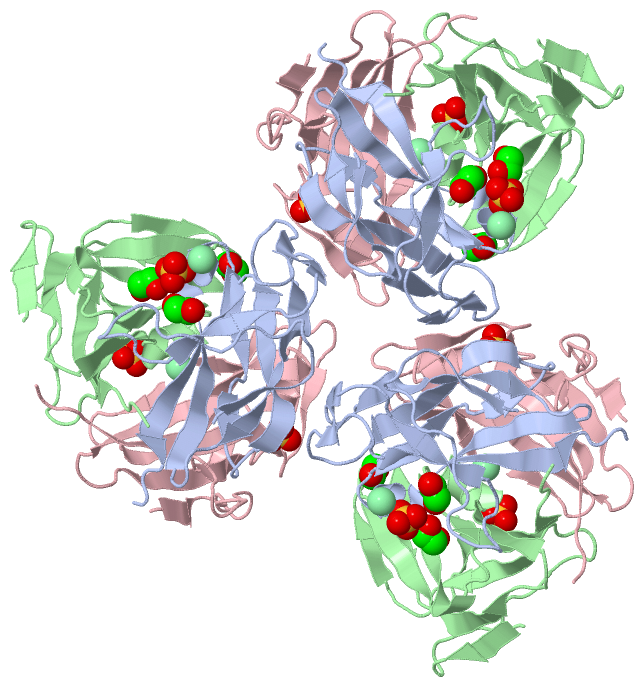
Chains, Units
Summary Information (see also Sequences/Alignments below) |
Ligands, Modified Residues, Ions (3, 8)
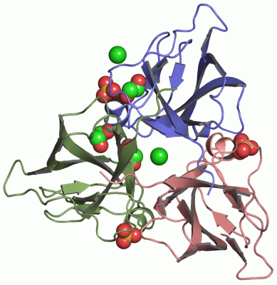
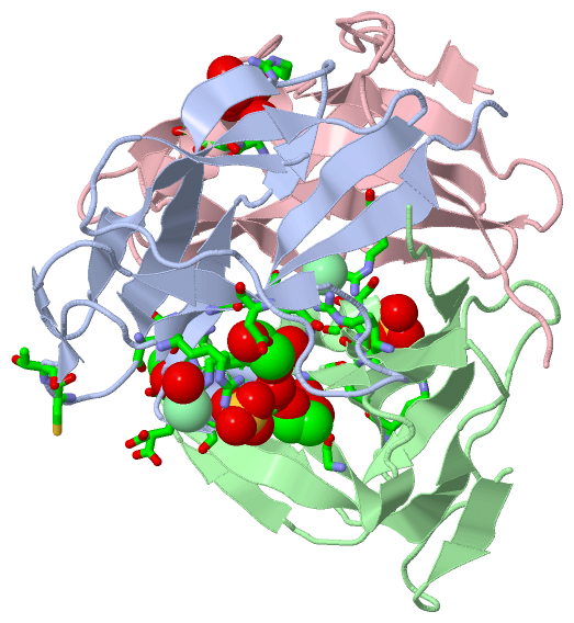
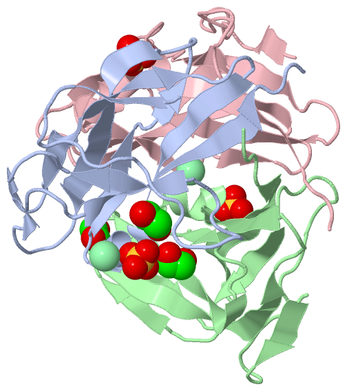
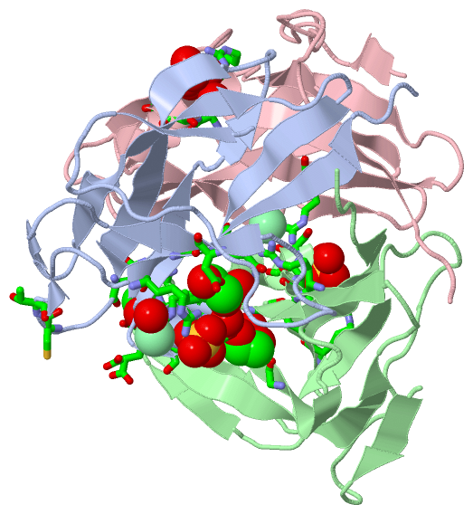
Asymmetric Unit (3, 8)
|
Sites (8, 8)
Asymmetric Unit (8, 8)
|
SS Bonds (0, 0)| (no "SS Bond" information available for 2OKB) |
Cis Peptide Bonds (0, 0)| (no "Cis Peptide Bond" information available for 2OKB) |
SAPs(SNPs)/Variants (0, 0)| (no "SAP(SNP)/Variant" information available for 2OKB) |
PROSITE Motifs (0, 0)| (no "PROSITE Motif" information available for 2OKB) |
Exons (0, 0)| (no "Exon" information available for 2OKB) |
Sequences/Alignments
Asymmetric UnitChain A from PDB Type:PROTEIN Length:113 aligned with A4GD96_9POXV | A4GD96 from UniProtKB/TrEMBL Length:147 Alignment length:113 20 30 40 50 60 70 80 90 100 110 120 A4GD96_9POXV 11 RFVKETNRAKSPTRQSPGAAGYDLYSAYDYTIPPGERQLIKTDISMSMPKFCYGRIAPRSGLSLKGIDIGGGVIDEDYRGNIGVILINNGKCTFNVNTGDRIAQLIYQRIYYP 123 SCOP domains d2okba_ A: Deoxyuridine 5'-triphosphate nucleotidohydrolase (dUTPase) SCOP domains CATH domains 2okbA00 A:11-123 [code=2.70.40.10, no name defined] CATH domains Pfam domains ----------------------------------------------------------------------------------------------------------------- Pfam domains SAPs(SNPs) ----------------------------------------------------------------------------------------------------------------- SAPs(SNPs) PROSITE ----------------------------------------------------------------------------------------------------------------- PROSITE Transcript ----------------------------------------------------------------------------------------------------------------- Transcript 2okb A 11 MFVKETNRAKSPTRQSPGAAGYDLYSAYDYTIPPGERQLIKTDISMSMPKFCYGRIAPRSGLSLKGIDIGGGVIDEDYRGNIGVILINNGKCTFNVNTGDRIAQLIYQRIYYP 123 20 30 40 50 60 70 80 90 100 110 120 Chain B from PDB Type:PROTEIN Length:113 aligned with A4GD96_9POXV | A4GD96 from UniProtKB/TrEMBL Length:147 Alignment length:113 20 30 40 50 60 70 80 90 100 110 120 A4GD96_9POXV 11 RFVKETNRAKSPTRQSPGAAGYDLYSAYDYTIPPGERQLIKTDISMSMPKFCYGRIAPRSGLSLKGIDIGGGVIDEDYRGNIGVILINNGKCTFNVNTGDRIAQLIYQRIYYP 123 SCOP domains d2okbb_ B: Deoxyuridine 5'-triphosphate nucleotidohydrolase (dUTPase) SCOP domains CATH domains 2okbB00 B:11-123 [code=2.70.40.10, no name defined] CATH domains Pfam domains ----------------------------------------------------------------------------------------------------------------- Pfam domains SAPs(SNPs) ----------------------------------------------------------------------------------------------------------------- SAPs(SNPs) PROSITE ----------------------------------------------------------------------------------------------------------------- PROSITE Transcript ----------------------------------------------------------------------------------------------------------------- Transcript 2okb B 11 MFVKETNRAKSPTRQSPGAAGYDLYSAYDYTIPPGERQLIKTDISMSMPKFCYGRIAPRSGLSLKGIDIGGGVIDEDYRGNIGVILINNGKCTFNVNTGDRIAQLIYQRIYYP 123 20 30 40 50 60 70 80 90 100 110 120 Chain C from PDB Type:PROTEIN Length:113 aligned with A4GD96_9POXV | A4GD96 from UniProtKB/TrEMBL Length:147 Alignment length:113 20 30 40 50 60 70 80 90 100 110 120 A4GD96_9POXV 11 RFVKETNRAKSPTRQSPGAAGYDLYSAYDYTIPPGERQLIKTDISMSMPKFCYGRIAPRSGLSLKGIDIGGGVIDEDYRGNIGVILINNGKCTFNVNTGDRIAQLIYQRIYYP 123 SCOP domains d2okbc_ C: Deoxyuridine 5'-triphosphate nucleotidohydrolase (dUTPase) SCOP domains CATH domains 2okbC00 C:11-123 [code=2.70.40.10, no name defined] CATH domains Pfam domains (1) ------dUTPase-2okbC01 C:17-123 Pfam domains (1) Pfam domains (2) ------dUTPase-2okbC02 C:17-123 Pfam domains (2) Pfam domains (3) ------dUTPase-2okbC03 C:17-123 Pfam domains (3) SAPs(SNPs) ----------------------------------------------------------------------------------------------------------------- SAPs(SNPs) PROSITE ----------------------------------------------------------------------------------------------------------------- PROSITE Transcript ----------------------------------------------------------------------------------------------------------------- Transcript 2okb C 11 MFVKETNRAKSPTRQSPGAAGYDLYSAYDYTIPPGERQLIKTDISMSMPKFCYGRIAPRSGLSLKGIDIGGGVIDEDYRGNIGVILINNGKCTFNVNTGDRIAQLIYQRIYYP 123 20 30 40 50 60 70 80 90 100 110 120
|
||||||||||||||||||||
SCOP Domains (1, 3)
Asymmetric Unit
 |
CATH Domains (1, 3)
Asymmetric Unit
|
Pfam Domains (1, 3)
Asymmetric Unit
|
Gene Ontology (3, 3)|
Asymmetric Unit(hide GO term definitions) Chain A,B,C (A4GD96_9POXV | A4GD96)
|
||||||||||||||||||||||||||||||
Interactive Views
|
||||||||||||||||||||||||||||||||||||||||||||||||||||||||||||||||||||||||||||||||||||||||||||||||||||||||||||||||||||||||||||||||||||||||||||||||||||||||||||||||||||||||||||||||||||||||||||||||||||||||||||
Still Images
|
||||||||||||||||
Databases
|
||||||||||||||||||||||||||||||||||||||||||||||||||||||||||||||||||||||||||||||||||||||||||||||||||||||||||||||||||||||||||||||||||||||||||||||||||||||||||||||||
Analysis Tools
|
|||||||||||||||||||||||||||||||||||||||||||||||||||||||||||||
Entries Sharing at Least One Protein Chain (UniProt ID)
Related Entries Specified in the PDB File
|
|