|
|
|
|
 Description
|
|
Compounds
|
||||||||||||||||||||||||||||||||||||||||||||||||||||
Chains, Units
Summary Information (see also Sequences/Alignments below) |
Ligands, Modified Residues, Ions (4, 7)| Asymmetric Unit (4, 7) Biological Unit 1 (3, 24) |
Sites (6, 6)
Asymmetric Unit (6, 6)
|
SS Bonds (0, 0)| (no "SS Bond" information available for 2OGG) |
Cis Peptide Bonds (2, 2)
Asymmetric Unit
|
||||||||||||
SAPs(SNPs)/Variants (0, 0)| (no "SAP(SNP)/Variant" information available for 2OGG) |
PROSITE Motifs (0, 0)| (no "PROSITE Motif" information available for 2OGG) |
Exons (0, 0)| (no "Exon" information available for 2OGG) |
Sequences/Alignments
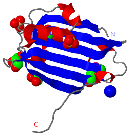
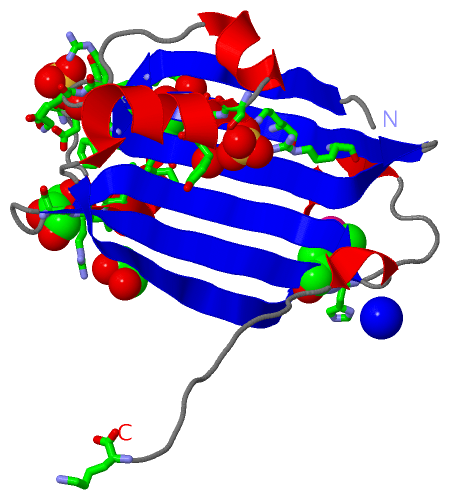
Asymmetric UnitChain A from PDB Type:PROTEIN Length:144 aligned with TRER_BACSU | P39796 from UniProtKB/Swiss-Prot Length:238 Alignment length:144 104 114 124 134 144 154 164 174 184 194 204 214 224 234 TRER_BACSU 95 TKTTVHKFGLEPPSELIQKQLRANLDDDIWEVIRSRKIDGEHVILDKDYFFRKHVPHLTKEICENSIYEYIEGELGLSISYAQKEIVAEPCTDEDRELLDLRGYDHMVVVRNYVFLEDTSLFQYTESRHRLDKFRFVDFARRGK 238 SCOP domains d2ogga1 A:95-238 Trehalose operon transcriptional repressor TreR SCOP domains CATH domains 2oggA01 A:95-228 Chorismate lyase ---------- CATH domains Pfam domains UTRA-2oggA01 A:95-229 --------- Pfam domains SAPs(SNPs) ------------------------------------------------------------------------------------------------------------------------------------------------ SAPs(SNPs) PROSITE ------------------------------------------------------------------------------------------------------------------------------------------------ PROSITE Transcript ------------------------------------------------------------------------------------------------------------------------------------------------ Transcript 2ogg A 95 TKTTVHKFGLEPPSELIQKQLRANLDDDIWEVIRSRKIDGEHVILDKDYFFRKHVPHLTKEICENSIYEYIEGELGLSISYAQKEIVAEPCTDEDRELLDLRGYDHmVVVRNYVFLEDTSLFQYTESRHRLDKFRFVDFARRGK 238 104 114 124 134 144 154 164 174 184 194 |204 214 224 234 201-MSE
|
||||||||||||||||||||
SCOP Domains (1, 1)
Asymmetric Unit
 |
CATH Domains (1, 1)
Asymmetric Unit
|
Pfam Domains (1, 1)| Asymmetric Unit |
Gene Ontology (4, 4)|
Asymmetric Unit(hide GO term definitions) Chain A (TRER_BACSU | P39796)
|
||||||||||||||||||||||||||||||||||||
Interactive Views
|
||||||||||||||||||||||||||||||||||||||||||||||||||||||||||||||||||||||||||||||||||||||||||||||||||||||||||||||||||||||||||||||||||||||||||||||||||||||||||||||||||||||||||||||||||||||||||||||||||||||||
Still Images
|
||||||||||||||||
Databases
|
||||||||||||||||||||||||||||||||||||||||||||||||||||||||||||||||||||||||||||||||||||||||||||||||||||||||||||||||||||||||||||||||||||||||||||||||||||||||||||||||
Analysis Tools
|
|||||||||||||||||||||||||||||||||||||||||||||||||||||||||||||
Entries Sharing at Least One Protein Chain (UniProt ID)
Related Entries Specified in the PDB File
|
|