|
|
|
|
 Description
|
|
Compounds
|
||||||||||||||||||||||||||||||||||||||||||||||||
Chains, Units
Summary Information (see also Sequences/Alignments below) |
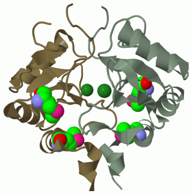
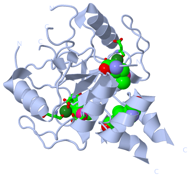
Ligands, Modified Residues, Ions (2, 3)| Asymmetric Unit (2, 3) Biological Unit 1 (1, 4) |
Sites (1, 1)
Asymmetric Unit (1, 1)
|
SS Bonds (0, 0)| (no "SS Bond" information available for 2OBB) |
Cis Peptide Bonds (1, 1)
Asymmetric Unit
|
||||||||
SAPs(SNPs)/Variants (0, 0)| (no "SAP(SNP)/Variant" information available for 2OBB) |
PROSITE Motifs (0, 0)| (no "PROSITE Motif" information available for 2OBB) |
Exons (0, 0)| (no "Exon" information available for 2OBB) |
Sequences/Alignments
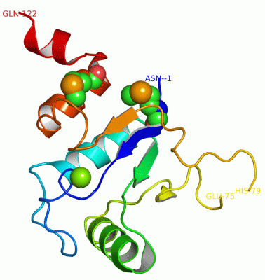
Asymmetric UnitChain A from PDB Type:PROTEIN Length:121 aligned with Q8A9J5_BACTN | Q8A9J5 from UniProtKB/TrEMBL Length:139 Alignment length:124 1 | 8 18 28 38 48 58 68 78 88 98 108 118 Q8A9J5_BACTN - --MTIAVDFDGTIVEHRYPRIGEEIPFAVETLKLLQQEKHRLILWSVREGELLDEAIEWCRARGLEFYAANKDYPEEERDHQGFSRKLKADLFIDDRNVGGIPDWGIIYEMIKEKKTFADIYSQ 122 SCOP domains --d2obba1 A:1-122 Hypothetical protein BT0820 SCOP domains CATH domains 2obbA00 A:-1-122 [code=3.40.50.1000, no name defined] CATH domains Pfam domains ---------------------------------------------------------------------------------------------------------------------------- Pfam domains SAPs(SNPs) ---------------------------------------------------------------------------------------------------------------------------- SAPs(SNPs) PROSITE ---------------------------------------------------------------------------------------------------------------------------- PROSITE Transcript ---------------------------------------------------------------------------------------------------------------------------- Transcript 2obb A -1 NAmTIAVDFDGTIVEHRYPRIGEEIPFAVETLKLLQQEKHRLILWSVREGELLDEAIEWCRARGLEFYAANKDYPEE---HQGFSRKLKADLFIDDRNVGGIPDWGIIYEmIKEKKTFADIYSQ 122 | 8 18 28 38 48 58 68 | -| 88 98 108| 118 | 75 79 109-MSE 1-MSE
|
||||||||||||||||||||
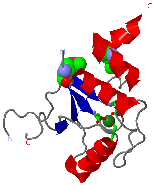
SCOP Domains (1, 1)
Asymmetric Unit
 |
CATH Domains (1, 1)
Asymmetric Unit
|
Pfam Domains (0, 0)| (no "Pfam Domain" information available for 2OBB) |
Gene Ontology (1, 1)|
Asymmetric Unit(hide GO term definitions) Chain A (Q8A9J5_BACTN | Q8A9J5)
|
||||||||||||
Interactive Views
|
||||||||||||||||||||||||||||||||||||||||||||||||||||||||||||||||||||||||||||||||||||||||||||||||||||||||||||||||||||||||||||||||||||||||||||||||
Still Images
|
||||||||||||||||
Databases
|
||||||||||||||||||||||||||||||||||||||||||||||||||||||||||||||||||||||||||||||||||||||||||||||||||||||||||||||||||||||||||||||||||||||||||||||||||||||||||||||||
Analysis Tools
|
|||||||||||||||||||||||||||||||||||||||||||||||||||||||||||||
Entries Sharing at Least One Protein Chain (UniProt ID)
Related Entries Specified in the PDB File
|
|