|
|
|
|
 Description
|
|
Compounds
|
||||||||||||||||||||||||||||||||||||||||||||||||||||||||
Chains, Units
Summary Information (see also Sequences/Alignments below) |
Ligands, Modified Residues, Ions (4, 7)| Asymmetric Unit (4, 7) Biological Unit 1 (3, 10) |
Sites (4, 4)
Asymmetric Unit (4, 4)
|
SS Bonds (0, 0)| (no "SS Bond" information available for 2OAI) |
Cis Peptide Bonds (0, 0)| (no "Cis Peptide Bond" information available for 2OAI) |
SAPs(SNPs)/Variants (0, 0)| (no "SAP(SNP)/Variant" information available for 2OAI) |
PROSITE Motifs (0, 0)| (no "PROSITE Motif" information available for 2OAI) |
Exons (0, 0)| (no "Exon" information available for 2OAI) |
Sequences/Alignments
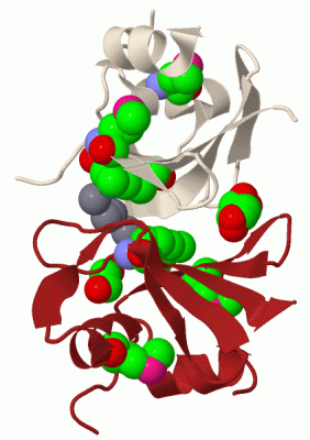
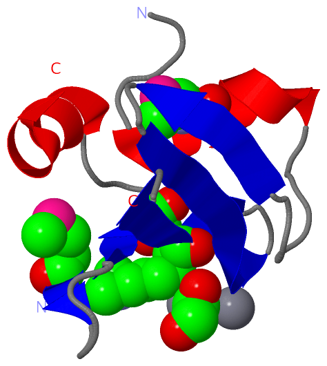
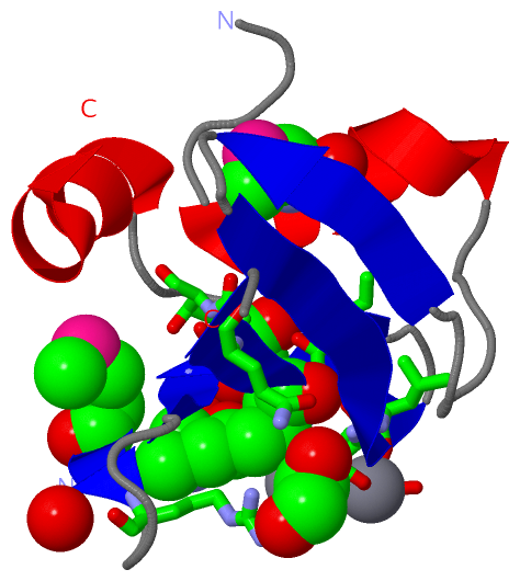
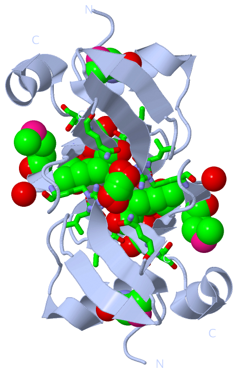
Asymmetric UnitChain A from PDB Type:PROTEIN Length:80 aligned with Q87DZ3_XYLFT | Q87DZ3 from UniProtKB/TrEMBL Length:448 Alignment length:87 362 372 382 392 402 412 422 432 Q87DZ3_XYLFT 353 EDALMVTREDGSFLIDGTLPIEELREVLGAELPDGEENNYHTLAGMCISYFGRIPHVGEYFDWAGWRIEIVDLDGARIDKLLLQRLN 439 SCOP domains d2oaia1 A:5-91 Hemolysin TlyC SCOP domains CATH domains 2oaiA00 A:5-91 [code=3.30.465 .10, no name defined] CATH domains Pfam domains -----CorC_HlyC-2oaiA01 A:10-91 Pfam domains SAPs(SNPs) --------------------------------------------------------------------------------------- SAPs(SNPs) PROSITE --------------------------------------------------------------------------------------- PROSITE Transcript --------------------------------------------------------------------------------------- Transcript 2oai A 5 EDALmVTREDGSFLIDGTLPIEELREVLGA-------NNYHTLAGmCISYFGRIPHVGEYFDWAGWRIEIVDLDGARIDkLLLQRLN 91 | 14 24 34 |44 | 54 64 74 84 | 34 42 50-MSE 84-MLY 9-MSE
|
||||||||||||||||||||
SCOP Domains (1, 1)
Asymmetric Unit
 |
CATH Domains (1, 1)
Asymmetric Unit
|
Pfam Domains (1, 1)
Asymmetric Unit
|
Gene Ontology (7, 7)|
Asymmetric Unit(hide GO term definitions) Chain A (Q87DZ3_XYLFT | Q87DZ3)
|
||||||||||||||||||||||||||||||||||||||||||||||||||||||||||||
Interactive Views
|
||||||||||||||||||||||||||||||||||||||||||||||||||||||||||||||||||||||||||||||||||||||||||||||||||||||||||||||||||||||||||||||||||||||||||||||||||||||||||||||||||||||||||||||||||
Still Images
|
||||||||||||||||
Databases
|
||||||||||||||||||||||||||||||||||||||||||||||||||||||||||||||||||||||||||||||||||||||||||||||||||||||||||||||||||||||||||||||||||||||||||||||||||||||||||||||||
Analysis Tools
|
|||||||||||||||||||||||||||||||||||||||||||||||||||||||||||||
Entries Sharing at Least One Protein Chain (UniProt ID)
Related Entries Specified in the PDB File
|
|