|
|
|
|
 Description
|
|
Compounds
|
||||||||||||||||||||||||||||||||||||||||||||||||
Chains, Units
Summary Information (see also Sequences/Alignments below) |
Ligands, Modified Residues, Ions (1, 1)
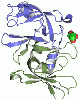
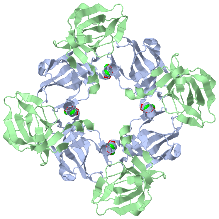
Asymmetric Unit (1, 1)
|
Sites (1, 1)
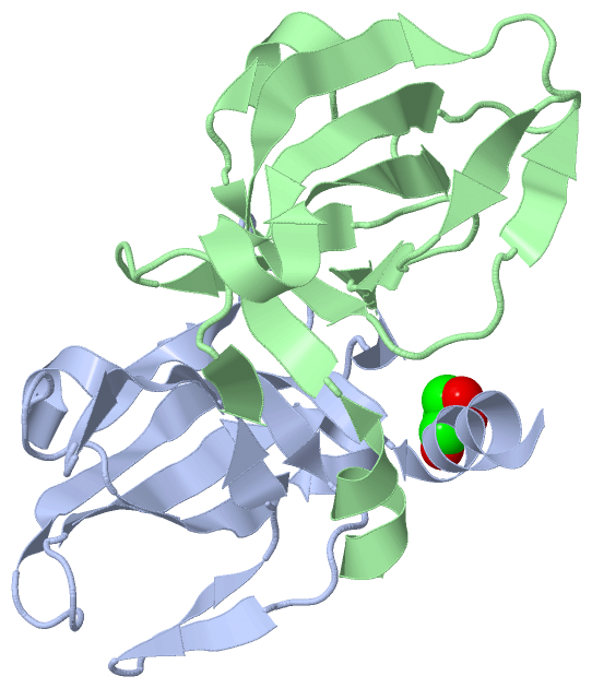
Asymmetric Unit (1, 1)
|
SS Bonds (0, 0)| (no "SS Bond" information available for 2NZH) |
Cis Peptide Bonds (0, 0)| (no "Cis Peptide Bond" information available for 2NZH) |
SAPs(SNPs)/Variants (0, 0)| (no "SAP(SNP)/Variant" information available for 2NZH) |
PROSITE Motifs (1, 2)
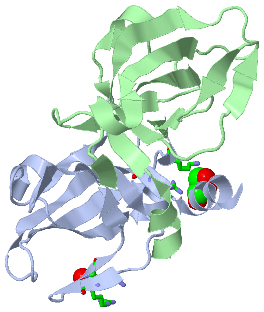
Asymmetric Unit (1, 2)
|
||||||||||||||||||||||||||||||||||||||||||||||||||||||||||||||||||||||||
Exons (0, 0)| (no "Exon" information available for 2NZH) |
Sequences/Alignments
Asymmetric UnitChain A from PDB Type:PROTEIN Length:110 aligned with CSAA_BACSU | P37584 from UniProtKB/Swiss-Prot Length:110 Alignment length:110 10 20 30 40 50 60 70 80 90 100 110 CSAA_BACSU 1 MAVIDDFEKLDIRTGTIVKAEEFPEARVPAIKLVIDFGTEIGIKQSSAQITKRYKPEGLINKQVIAVVNFPPRRIAGFKSEVLVLGGIPGQGDVVLLQPDQPVPNGTKIG 110 SCOP domains d2nzha_ A: automated matches SCOP domains CATH domains 2nzhA00 A:1-110 Nucleic acid-binding proteins CATH domains Pfam domains -------------------------------------------------------------------------------------------------------------- Pfam domains SAPs(SNPs) -------------------------------------------------------------------------------------------------------------- SAPs(SNPs) PROSITE -----TRBD PDB: A:6-110 UniProt: 6-110 PROSITE Transcript -------------------------------------------------------------------------------------------------------------- Transcript 2nzh A 1 MAVIDDFEKLDIRTGTIVKAEEFPEARVPAIKLVIDFGTEIGIKQSSAQITKRYKPEGLINKQVIAVVNFPPRRIAGFKSEVLVLGGIPGQGDVVLLQPDQPVPNGTKIG 110 10 20 30 40 50 60 70 80 90 100 110 Chain B from PDB Type:PROTEIN Length:108 aligned with CSAA_BACSU | P37584 from UniProtKB/Swiss-Prot Length:110 Alignment length:108 12 22 32 42 52 62 72 82 92 102 CSAA_BACSU 3 VIDDFEKLDIRTGTIVKAEEFPEARVPAIKLVIDFGTEIGIKQSSAQITKRYKPEGLINKQVIAVVNFPPRRIAGFKSEVLVLGGIPGQGDVVLLQPDQPVPNGTKIG 110 SCOP domains d2nzhb_ B: automated matches SCOP domains CATH domains 2nzhB00 B:3-110 Nucleic acid-binding proteins CATH domains Pfam domains (1) ---------tRNA_bind-2nzhB01 B:12-108 -- Pfam domains (1) Pfam domains (2) ---------tRNA_bind-2nzhB02 B:12-108 -- Pfam domains (2) SAPs(SNPs) ------------------------------------------------------------------------------------------------------------ SAPs(SNPs) PROSITE ---TRBD PDB: B:6-110 UniProt: 6-110 PROSITE Transcript ------------------------------------------------------------------------------------------------------------ Transcript 2nzh B 3 VIDDFEKLDIRTGTIVKAEEFPEARVPAIKLVIDFGTEIGIKQSSAQITKRYKPEGLINKQVIAVVNFPPRRIAGFKSEVLVLGGIPGQGDVVLLQPDQPVPNGTKIG 110 12 22 32 42 52 62 72 82 92 102
|
||||||||||||||||||||
SCOP Domains (1, 2)
Asymmetric Unit
 |
CATH Domains (1, 2)
Asymmetric Unit
|
Pfam Domains (1, 2)
Asymmetric Unit
|
Gene Ontology (5, 5)|
Asymmetric Unit(hide GO term definitions) Chain A,B (CSAA_BACSU | P37584)
|
||||||||||||||||||||||||||||||||||||||||||||||||
Interactive Views
|
|||||||||||||||||||||||||||||||||||||||||||||||||||||||||||||||||||||||||||||||||||||||||||||||||||||||||||||||||||||||||||||||||||||||||||||
Still Images
|
||||||||||||||||
Databases
|
||||||||||||||||||||||||||||||||||||||||||||||||||||||||||||||||||||||||||||||||||||||||||||||||||||||||||||||||||||||||||||||||||||||||||||||||||||||||||||||||
Analysis Tools
|
|||||||||||||||||||||||||||||||||||||||||||||||||||||||||||||
Entries Sharing at Least One Protein Chain (UniProt ID)
Related Entries Specified in the PDB File
|
|