|
|
|
|
 Description
|
|
Compounds
|
||||||||||||||||||||||||||||||||||||||||||||||||
Chains, Units
Summary Information (see also Sequences/Alignments below) |
Ligands, Modified Residues, Ions (0, 0)| (no "Ligand,Modified Residues,Ions" information available for 2MGV) |
Sites (0, 0)| (no "Site" information available for 2MGV) |
SS Bonds (0, 0)| (no "SS Bond" information available for 2MGV) |
Cis Peptide Bonds (0, 0)| (no "Cis Peptide Bond" information available for 2MGV) |
SAPs(SNPs)/Variants (0, 0)| (no "SAP(SNP)/Variant" information available for 2MGV) |
PROSITE Motifs (0, 0)| (no "PROSITE Motif" information available for 2MGV) |
Exons (0, 0)| (no "Exon" information available for 2MGV) |
Sequences/Alignments
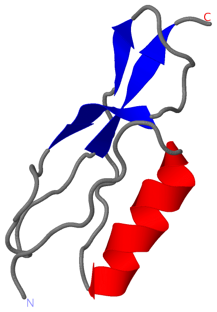
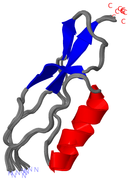
NMR StructureChain A from PDB Type:PROTEIN Length:65 aligned with I6YGX2_MYCTU | I6YGX2 from UniProtKB/TrEMBL Length:810 Alignment length:65 709 719 729 739 749 759 I6YGX2_MYCTU 700 GSRVPSVAGLDVDAARQRLKDAGFQVADQTNSVNSSAKYGEVVGTSPSGQTIPGSIVTIQISNGI 764 SCOP domains ----------------------------------------------------------------- SCOP domains CATH domains ----------------------------------------------------------------- CATH domains Pfam domains ----------------------------------------------------------------- Pfam domains SAPs(SNPs) ----------------------------------------------------------------- SAPs(SNPs) PROSITE ----------------------------------------------------------------- PROSITE Transcript ----------------------------------------------------------------- Transcript 2mgv A 1 GSRVPSVAGLDVDAARQRLKDAGFQVADQTNSVNSSAKYGEVVGTSPSGQTIPGSIVTIQISNGI 65 10 20 30 40 50 60
|
||||||||||||||||||||
SCOP Domains (0, 0)| (no "SCOP Domain" information available for 2MGV) |
CATH Domains (0, 0)| (no "CATH Domain" information available for 2MGV) |
Pfam Domains (0, 0)| (no "Pfam Domain" information available for 2MGV) |
Gene Ontology (1, 1)|
NMR Structure(hide GO term definitions) Chain A (I6YGX2_MYCTU | I6YGX2)
|
||||||||||||
Interactive Views
|
||||||||||||||||||||||||||||||||||||||||||||||||||||||||||||||||||||||||||||||||||||||||||||||||||||||||||||||||||||
Still Images
|
||||||||||||||||
Databases
|
||||||||||||||||||||||||||||||||||||||||||||||||||||||||||||||||||||||||||||||||||||||||||||||||||||||||||||||||||||||||||||||||||||||||||||||||||||||||||||||||
Analysis Tools
|
|||||||||||||||||||||||||||||||||||||||||||||||||||||||||||||
Entries Sharing at Least One Protein Chain (UniProt ID)
Related Entries Specified in the PDB File
|
|