Show PDB file:   
         Plain Text   HTML   (compressed file size)
QuickSearch:   
by PDB,NDB,UniProt,PROSITE Code or Search Term(s)  
(-)NMR Structure - model 1
(-)NMR Structure - model 1, sites
(-)NMR Structure - all models
collapse expand < >
Image NMR Structure - model 1
NMR Structure - model 1  (Jmol Viewer)
Image NMR Structure - model 1, sites
NMR Structure - model 1, sites  (Jmol Viewer)
Image NMR Structure - all models
NMR Structure - all models  (Jmol Viewer)

(-) Description

Title :  NMR STRUCTURE OF SP140 PHD FINGER CIS CONFORMER
 
Authors :  C. Zucchelli, G. Quilici, G. Musco
Date :  02 Sep 13  (Deposition) - 13 Nov 13  (Release) - 22 Jan 14  (Revision)
Method :  SOLUTION NMR
Resolution :  NOT APPLICABLE
Chains :  NMR Structure  :  C  (20x)
NMR Structure *:  C  (1x)
Keywords :  Phd Finger, Transcription (Keyword Search: [Gene Ontology, PubMed, Web (Google))
 
Reference :  C. Zucchelli, S. Tamburri, G. Quilici, E. Palagano, A. Berardi, M. Saare, P. Peterson, A. Bachi, G. Musco
Structure Of Human Sp140 Phd Finger: An Atypical Fold Interacting With Pin1.
Febs J. V. 281 216 2014
PubMed-ID: 24267382  |  Reference-DOI: 10.1111/FEBS.12588

(-) Compounds

Molecule 1 - NUCLEAR BODY PROTEIN SP140
    ChainsC
    EngineeredYES
    Expression SystemESCHERICHIA COLI
    Expression System PlasmidPETM11
    Expression System StrainBL21(DE3)
    Expression System Taxid469008
    Expression System Vector TypePLASMID
    FragmentUNP RESIDUES 687-738
    Organism CommonHUMAN
    Organism ScientificHOMO SAPIENS
    Organism Taxid9606
    SynonymLYMPHOID-RESTRICTED HOMOLOG OF SP100, LYSP100, NUCLEAR AUTOANTIGEN SP-140, SPECKLED 140 KDA

 Structural Features

(-) Chains, Units

  1
NMR Structure (20x)C
NMR Structure * (1x)C

Summary Information (see also Sequences/Alignments below)

(-) Ligands, Modified Residues, Ions  (1, 2)

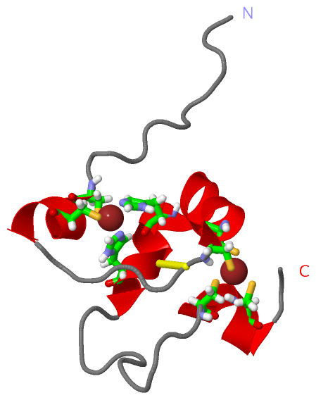
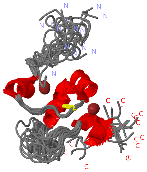
NMR Structure (1, 2)
No.NameCountTypeFull Name
1ZN2Ligand/IonZINC ION
NMR Structure * (0, 0)
No.NameCountTypeFull Name
1ZN-1Ligand/IonZINC ION

(-) Sites  (2, 2)

NMR Structure (2, 2)
No.NameEvidenceResiduesDescription
1AC1SOFTWARECYS C:23 , CYS C:26 , CYS C:48 , CYS C:51BINDING SITE FOR RESIDUE ZN C 101
2AC2SOFTWARECYS C:11 , CYS C:14 , HIS C:31 , HIS C:35BINDING SITE FOR RESIDUE ZN C 102

(-) SS Bonds  (1, 1)

NMR Structure
No.Residues
1C:22 -C:34

(-) Cis Peptide Bonds  (2, 40)

NMR Structure
No.ModelResidues
11, 2, 3, 4, 5, 6, 7, 8, 9, 10, 11, 12, 13, 14, 15, 16, 17, 18, 19, 20Ile C:36 -Pro C:37
21, 2, 3, 4, 5, 6, 7, 8, 9, 10, 11, 12, 13, 14, 15, 16, 17, 18, 19, 20Thr C:44 -Pro C:45

 Sequence-Structure Mapping

(-) SAPs(SNPs)/Variants  (0, 0)

(no "SAP(SNP)/Variant" information available for 2MD8)

(-) PROSITE Motifs  (2, 2)

NMR Structure (2, 2)
 PROSITEUniProtKBPDB
No.IDACDescriptionIDLocationCountLocation
1ZF_PHD_2PS50016 Zinc finger PHD-type profile.SP140_HUMAN690-736  1C:8-54
2ZF_PHD_1PS01359 Zinc finger PHD-type signature.SP140_HUMAN693-733  1C:11-51
NMR Structure * (2, 2)
 PROSITEUniProtKBPDB
No.IDACDescriptionIDLocationCountLocation
1ZF_PHD_2PS50016 Zinc finger PHD-type profile.SP140_HUMAN690-736  1C:8-54
2ZF_PHD_1PS01359 Zinc finger PHD-type signature.SP140_HUMAN693-733  1C:11-51

(-) Exons   (0, 0)

(no "Exon" information available for 2MD8)

(-) Sequences/Alignments

NMR Structure
   Reformat: Number of residues per line =  ('0' or empty: single-line sequence representation)
  Number of residues per labelling interval =   
  UniProt sequence: complete  aligned part    
   Show mapping: SCOP domains CATH domains Pfam domains Secondary structure (by author)
SAPs(SNPs) PROSITE motifs Exons
(details for a mapped element are shown in a popup box when the mouse pointer rests over it)
Chain C from PDB  Type:PROTEIN  Length:56
 aligned with SP140_HUMAN | Q13342 from UniProtKB/Swiss-Prot  Length:867

    Alignment length:91
                                   657       667       677       687       697       707       717       727       737 
          SP140_HUMAN   648 GWPLRWLMENGFLPDPPRIRYRKKKRILKSQNNSSVDPCMRNLDECEVCRDGGELFCCDTCSRVFHEDCHIPPVEAERTPWNCIFCRMKES 738
               SCOP domains d     2md                              8c_ C: automated matches                             SCOP domains
               CATH domains ------------------------------------------------------------------------------------------- CATH domains
               Pfam domains ------------------------------------------------------------------------------------------- Pfam domains
         Sec.struct. author .-----...------------------------------......hhhhhh......hhhhhhhhhhhhhh...........hhhhhh... Sec.struct. author
                 SAPs(SNPs) ------------------------------------------------------------------------------------------- SAPs(SNPs)
                PROSITE (1) ------------------------------------------ZF_PHD_2  PDB: C:8-54 UniProt: 690-736         -- PROSITE (1)
                PROSITE (2) ---------------------------------------------ZF_PHD_1  PDB: C:11-51 UniProt: 693-733  ----- PROSITE (2)
                 Transcript ------------------------------------------------------------------------------------------- Transcript
                 2md8 C   1 G-----AMG------------------------------MRNLDECEVCRDGGELFCCDTCSRVFHEDCHIPPVEAERTPWNCIFCRMKES  56
                            |     | |-         -         -         5        15        25        35        45        55 
                            |     2 4                              5                                                   
                            1                                                                                          

   Legend:   → Mismatch (orange background)
  - → Gap (green background, '-', border residues have a numbering label)
    → Modified Residue (blue background, lower-case, 'x' indicates undefined single-letter code, labelled with number + name)
  x → Chemical Group (purple background, 'x', labelled with number + name, e.g. ACE or NH2)
  extra numbering lines below/above indicate numbering irregularities and modified residue names etc., number ends below/above '|'

 Classification and Annotation

(-) SCOP Domains  (1, 1)

NMR Structure

(-) CATH Domains  (0, 0)

(no "CATH Domain" information available for 2MD8)

(-) Pfam Domains  (0, 0)

(no "Pfam Domain" information available for 2MD8)

(-) Gene Ontology  (14, 14)

NMR Structure(hide GO term definitions)
Chain C   (SP140_HUMAN | Q13342)
molecular function
    GO:0003677    DNA binding    Any molecular function by which a gene product interacts selectively and non-covalently with DNA (deoxyribonucleic acid).
    GO:0046872    metal ion binding    Interacting selectively and non-covalently with any metal ion.
    GO:0005515    protein binding    Interacting selectively and non-covalently with any protein or protein complex (a complex of two or more proteins that may include other nonprotein molecules).
    GO:0003700    transcription factor activity, sequence-specific DNA binding    Interacting selectively and non-covalently with a specific DNA sequence in order to modulate transcription. The transcription factor may or may not also interact selectively with a protein or macromolecular complex.
    GO:0008270    zinc ion binding    Interacting selectively and non-covalently with zinc (Zn) ions.
biological process
    GO:0006952    defense response    Reactions, triggered in response to the presence of a foreign body or the occurrence of an injury, which result in restriction of damage to the organism attacked or prevention/recovery from the infection caused by the attack.
    GO:0006355    regulation of transcription, DNA-templated    Any process that modulates the frequency, rate or extent of cellular DNA-templated transcription.
cellular component
    GO:0016605    PML body    A class of nuclear body; they react against SP100 auto-antibodies (PML, promyelocytic leukemia); cells typically contain 10-30 PML bodies per nucleus; alterations in the localization of PML bodies occurs after viral infection.
    GO:0005737    cytoplasm    All of the contents of a cell excluding the plasma membrane and nucleus, but including other subcellular structures.
    GO:0005739    mitochondrion    A semiautonomous, self replicating organelle that occurs in varying numbers, shapes, and sizes in the cytoplasm of virtually all eukaryotic cells. It is notably the site of tissue respiration.
    GO:0005635    nuclear envelope    The double lipid bilayer enclosing the nucleus and separating its contents from the rest of the cytoplasm; includes the intermembrane space, a gap of width 20-40 nm (also called the perinuclear space).
    GO:0005730    nucleolus    A small, dense body one or more of which are present in the nucleus of eukaryotic cells. It is rich in RNA and protein, is not bounded by a limiting membrane, and is not seen during mitosis. Its prime function is the transcription of the nucleolar DNA into 45S ribosomal-precursor RNA, the processing of this RNA into 5.8S, 18S, and 28S components of ribosomal RNA, and the association of these components with 5S RNA and proteins synthesized outside the nucleolus. This association results in the formation of ribonucleoprotein precursors; these pass into the cytoplasm and mature into the 40S and 60S subunits of the ribosome.
    GO:0005654    nucleoplasm    That part of the nuclear content other than the chromosomes or the nucleolus.
    GO:0005634    nucleus    A membrane-bounded organelle of eukaryotic cells in which chromosomes are housed and replicated. In most cells, the nucleus contains all of the cell's chromosomes except the organellar chromosomes, and is the site of RNA synthesis and processing. In some species, or in specialized cell types, RNA metabolism or DNA replication may be absent.

 Visualization

(-) Interactive Views

NMR Structure
  Complete Structure
    Jena3D(integrated viewing of ligand, site, SAP, PROSITE, SCOP information)
    WebMol | AstexViewer[tm]@PDBe
(Java Applets, require no local installation except for Java; loading may be slow)
    STRAP
(Java WebStart application, automatic local installation, requires Java; full application with system access!)
    RasMol
(require local installation)
    Molscript (VRML)
(requires installation of a VRML viewer; select preferred view via VRML and generate a mono or stereo PDF format file)
 
  Ligands, Modified Residues, Ions
    ZN  [ RasMol | Jena3D ]  +environment [ RasMol | Jena3D ]
 
  Sites
    AC1  [ RasMol ]  +environment [ RasMol ]
    AC2  [ RasMol ]  +environment [ RasMol ]
 
  Cis Peptide Bonds
    Ile C:36 - Pro C:37   [ RasMol ]  
    Thr C:44 - Pro C:45   [ RasMol ]  
 

(-) Still Images

Jmol
  protein: cartoon or spacefill or dots and stick; nucleic acid: cartoon and stick; ligands: spacefill; active site: stick
Molscript
  protein, nucleic acid: cartoon; ligands: spacefill; active site: ball and stick

 Databases and Analysis Tools

(-) Databases

Access by PDB/NDB ID
  2md8
    Family and Domain InformationProDom | SYSTERS
    General Structural InformationGlycoscienceDB | MMDB | NDB | OCA | PDB | PDBe | PDBj | PDBsum | PDBWiki | PQS | PROTEOPEDIA
    Orientation in MembranesOPM
    Protein SurfaceSURFACE
    Secondary StructureDSSP (structure derived) | HSSP (homology derived)
    Structural GenomicsGeneCensus
    Structural NeighboursCE | VAST
    Structure ClassificationCATH | Dali | SCOP
    Validation and Original DataBMRB Data View | BMRB Restraints Grid | EDS | PROCHECK | RECOORD | WHAT_CHECK
 
Access by UniProt ID/Accession number
  SP140_HUMAN | Q13342
    Comparative Protein Structure ModelsModBase
    Genomic InformationEnsembl
    Protein-protein InteractionDIP
    Sequence, Family and Domain InformationInterPro | Pfam | SMART | UniProtKB/SwissProt
 
Access by Enzyme Classificator   (EC Number)
  (no 'Enzyme Classificator' available)
    General Enzyme InformationBRENDA | EC-PDB | Enzyme | IntEnz
    PathwayKEGG | MetaCyc
 
Access by Disease Identifier   (MIM ID)
  (no 'MIM ID' available)
    Disease InformationOMIM
 
Access by GenAge ID
  (no 'GenAge ID' available)
    Age Related InformationGenAge

(-) Analysis Tools

Access by PDB/NDB ID
    Domain InformationXDom
    Interatomic Contacts of Structural UnitsCSU
    Ligand-protein ContactsLPC
    Protein CavitiescastP
    Sequence and Secondary StructurePDBCartoon
    Structure AlignmentSTRAP(Java WebStart application, automatic local installation, requires Java; full application with system access!)
    Structure and Sequence BrowserSTING
 
Access by UniProt ID/Accession number
  SP140_HUMAN | Q13342
    Protein Disorder PredictionDisEMBL | FoldIndex | GLOBPLOT (for more information see DisProt)

 Related Entries

(-) Entries Sharing at Least One Protein Chain (UniProt ID)

UniProtKB/Swiss-Prot
        SP140_HUMAN | Q133422md7

(-) Related Entries Specified in the PDB File

2md7 RELATED ID: 19473 RELATED DB: BMRB