Show PDB file:   
         Plain Text   HTML   (compressed file size)
QuickSearch:   
by PDB,NDB,UniProt,PROSITE Code or Search Term(s)  
(-)NMR Structure - model 1
(-)NMR Structure - model 1, sites
(-)NMR Structure - all models
collapse expand < >
Image NMR Structure - model 1
NMR Structure - model 1  (Jmol Viewer)
Image NMR Structure - model 1, sites
NMR Structure - model 1, sites  (Jmol Viewer)
Image NMR Structure - all models
NMR Structure - all models  (Jmol Viewer)

(-) Description

Title :  SOLUTION NMR STRUCTURE OF FULL-LENGTH OXIDIZED MICROSOMAL RABBIT CYTOCHROME B5
 
Authors :  V. Subramanian, S. Ahuja, N. Popovych, R. Huang, S. V. Le Clair, N. Jahr J. Xu, K. Yamamoto, R. P. Nanga, S. Im, L. Waskell, A. Ramamoorthy
Date :  08 Jan 13  (Deposition) - 06 Feb 13  (Release) - 27 Apr 16  (Revision)
Method :  SOLUTION NMR
Resolution :  NOT APPLICABLE
Chains :  NMR Structure  :  A  (23x)
NMR Structure *:  A  (1x)
Keywords :  Membrane Protein, Heme, Haddock, Electron Transport (Keyword Search: [Gene Ontology, PubMed, Web (Google))
 
Reference :  S. Ahuja, V. Subramanian, N. Popovych, R. Huang, S. V. Le Clair, N. Jahr, R. Soong, J. Xu, K. Yamamoto, R. P. Nanga, S. Im, L. Waskell, A. Ramamoorthy
Nmr Structure Of Full-Length Mammalian Cytochrome B5
To Be Published
PubMed: search

(-) Compounds

Molecule 1 - CYTOCHROME B5
    ChainsA
    EngineeredYES
    Expression SystemESCHERICHIA COLI
    Expression System PlasmidPSC
    Expression System Taxid562
    Expression System Vector TypePLASMID
    FragmentUNP RESIDUES 1-104
    GeneCYB5A, CYB5
    Organism CommonRABBIT
    Organism ScientificORYCTOLAGUS CUNICULUS
    Organism Taxid9986

 Structural Features

(-) Chains, Units

  1
NMR Structure (23x)A
NMR Structure * (1x)A

Summary Information (see also Sequences/Alignments below)

(-) Ligands, Modified Residues, Ions  (1, 1)

NMR Structure (1, 1)
No.NameCountTypeFull Name
1HEM1Ligand/IonPROTOPORPHYRIN IX CONTAINING FE
NMR Structure * (1, 1)
No.NameCountTypeFull Name
1HEM1Ligand/IonPROTOPORPHYRIN IX CONTAINING FE

(-) Sites  (1, 1)

NMR Structure (1, 1)
No.NameEvidenceResiduesDescription
1AC1SOFTWARELEU A:30 , LEU A:37 , HIS A:44 , PRO A:45 , VAL A:50 , VAL A:66 , HIS A:68 , SER A:69BINDING SITE FOR RESIDUE HEM A 400

(-) SS Bonds  (0, 0)

(no "SS Bond" information available for 2M33)

(-) Cis Peptide Bonds  (0, 0)

(no "Cis Peptide Bond" information available for 2M33)

 Sequence-Structure Mapping

(-) SAPs(SNPs)/Variants  (0, 0)

(no "SAP(SNP)/Variant" information available for 2M33)

(-) PROSITE Motifs  (2, 2)

NMR Structure (2, 2)
 PROSITEUniProtKBPDB
No.IDACDescriptionIDLocationCountLocation
1CYTOCHROME_B5_2PS50255 Cytochrome b5 family, heme-binding domain profile.CYB5_RABIT9-85  1A:9-85
2CYTOCHROME_B5_1PS00191 Cytochrome b5 family, heme-binding domain signature.CYB5_RABIT40-47  1A:40-47
NMR Structure * (2, 2)
 PROSITEUniProtKBPDB
No.IDACDescriptionIDLocationCountLocation
1CYTOCHROME_B5_2PS50255 Cytochrome b5 family, heme-binding domain profile.CYB5_RABIT9-85  1A:9-85
2CYTOCHROME_B5_1PS00191 Cytochrome b5 family, heme-binding domain signature.CYB5_RABIT40-47  1A:40-47

(-) Exons   (0, 0)

(no "Exon" information available for 2M33)

(-) Sequences/Alignments

NMR Structure
   Reformat: Number of residues per line =  ('0' or empty: single-line sequence representation)
  Number of residues per labelling interval =   
  UniProt sequence: complete  aligned part    
   Show mapping: SCOP domains CATH domains Pfam domains Secondary structure (by author)
SAPs(SNPs) PROSITE motifs Exons
(details for a mapped element are shown in a popup box when the mouse pointer rests over it)
Chain A from PDB  Type:PROTEIN  Length:104
 aligned with CYB5_RABIT | P00169 from UniProtKB/Swiss-Prot  Length:134

    Alignment length:104
                                    10        20        30        40        50        60        70        80        90       100    
           CYB5_RABIT     1 MAAQSDKDVKYYTLEEIKKHNHSKSTWLILHHKVYDLTKFLEEHPGGEEVLREQAGGDATENFEDVGHSTDARELSKTFIIGELHPDDRSKLSKPMETLITTVD 104
               SCOP domains d2m33a_ A: automated matches                                                                             SCOP domains
               CATH domains -------------------------------------------------------------------------------------------------------- CATH domains
               Pfam domains -------------------------------------------------------------------------------------------------------- Pfam domains
         Sec.struct. author .........eeehhhhhh........eeee..eeee...hhhhh...hhhhhh.....hhhhhhhhh.hhhhhhhh...eeeee.hhhh............... Sec.struct. author
                 SAPs(SNPs) -------------------------------------------------------------------------------------------------------- SAPs(SNPs)
                PROSITE (1) --------CYTOCHROME_B5_2  PDB: A:9-85 UniProt: 9-85                                   ------------------- PROSITE (1)
                PROSITE (2) ---------------------------------------CYTOCHRO--------------------------------------------------------- PROSITE (2)
                 Transcript -------------------------------------------------------------------------------------------------------- Transcript
                 2m33 A   1 MAAQSDKDVKYYTLEEIKKHNHSKSTWLILHHKVYDLTKFLEEHPGGEEVLREQAGGDATENFEDVGHSTDARELSKTFIIGELHPDDRSKLSKPMETLITTVD 104
                                    10        20        30        40        50        60        70        80        90       100    

   Legend:   → Mismatch (orange background)
  - → Gap (green background, '-', border residues have a numbering label)
    → Modified Residue (blue background, lower-case, 'x' indicates undefined single-letter code, labelled with number + name)
  x → Chemical Group (purple background, 'x', labelled with number + name, e.g. ACE or NH2)
  extra numbering lines below/above indicate numbering irregularities and modified residue names etc., number ends below/above '|'

 Classification and Annotation

(-) SCOP Domains  (1, 1)

NMR Structure

(-) CATH Domains  (0, 0)

(no "CATH Domain" information available for 2M33)

(-) Pfam Domains  (0, 0)

(no "Pfam Domain" information available for 2M33)

(-) Gene Ontology  (13, 13)

NMR Structure(hide GO term definitions)
Chain A   (CYB5_RABIT | P00169)
molecular function
    GO:0019899    enzyme binding    Interacting selectively and non-covalently with any enzyme.
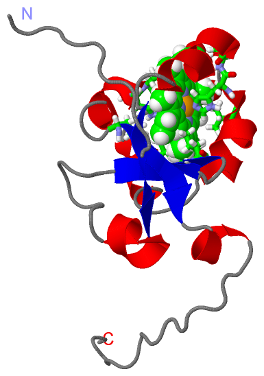
    GO:0020037    heme binding    Interacting selectively and non-covalently with heme, any compound of iron complexed in a porphyrin (tetrapyrrole) ring.
    GO:0046872    metal ion binding    Interacting selectively and non-covalently with any metal ion.
biological process
    GO:0055114    oxidation-reduction process    A metabolic process that results in the removal or addition of one or more electrons to or from a substance, with or without the concomitant removal or addition of a proton or protons.
cellular component
    GO:0005737    cytoplasm    All of the contents of a cell excluding the plasma membrane and nucleus, but including other subcellular structures.
    GO:0005783    endoplasmic reticulum    The irregular network of unit membranes, visible only by electron microscopy, that occurs in the cytoplasm of many eukaryotic cells. The membranes form a complex meshwork of tubular channels, which are often expanded into slitlike cavities called cisternae. The ER takes two forms, rough (or granular), with ribosomes adhering to the outer surface, and smooth (with no ribosomes attached).
    GO:0005789    endoplasmic reticulum membrane    The lipid bilayer surrounding the endoplasmic reticulum.
    GO:0070062    extracellular exosome    A vesicle that is released into the extracellular region by fusion of the limiting endosomal membrane of a multivesicular body with the plasma membrane. Extracellular exosomes, also simply called exosomes, have a diameter of about 40-100 nm.
    GO:0016021    integral component of membrane    The component of a membrane consisting of the gene products and protein complexes having at least some part of their peptide sequence embedded in the hydrophobic region of the membrane.
    GO:0043231    intracellular membrane-bounded organelle    Organized structure of distinctive morphology and function, bounded by a single or double lipid bilayer membrane and occurring within the cell. Includes the nucleus, mitochondria, plastids, vacuoles, and vesicles. Excludes the plasma membrane.
    GO:0016020    membrane    A lipid bilayer along with all the proteins and protein complexes embedded in it an attached to it.
    GO:0005739    mitochondrion    A semiautonomous, self replicating organelle that occurs in varying numbers, shapes, and sizes in the cytoplasm of virtually all eukaryotic cells. It is notably the site of tissue respiration.
    GO:0031090    organelle membrane    A membrane that is one of the two lipid bilayers of an organelle envelope or the outermost membrane of single membrane bound organelle.

 Visualization

(-) Interactive Views

NMR Structure
  Complete Structure
    Jena3D(integrated viewing of ligand, site, SAP, PROSITE, SCOP information)
    WebMol | AstexViewer[tm]@PDBe
(Java Applets, require no local installation except for Java; loading may be slow)
    STRAP
(Java WebStart application, automatic local installation, requires Java; full application with system access!)
    RasMol
(require local installation)
    Molscript (VRML)
(requires installation of a VRML viewer; select preferred view via VRML and generate a mono or stereo PDF format file)
 
  Ligands, Modified Residues, Ions
    HEM  [ RasMol | Jena3D ]  +environment [ RasMol | Jena3D ]
 
  Sites
    AC1  [ RasMol ]  +environment [ RasMol ]
 
  Cis Peptide Bonds
(no "Cis Peptide Bonds" information available for 2m33)
 

(-) Still Images

Jmol
  protein: cartoon or spacefill or dots and stick; nucleic acid: cartoon and stick; ligands: spacefill; active site: stick
Molscript
  protein, nucleic acid: cartoon; ligands: spacefill; active site: ball and stick

 Databases and Analysis Tools

(-) Databases

Access by PDB/NDB ID
  2m33
    Family and Domain InformationProDom | SYSTERS
    General Structural InformationGlycoscienceDB | MMDB | NDB | OCA | PDB | PDBe | PDBj | PDBsum | PDBWiki | PQS | PROTEOPEDIA
    Orientation in MembranesOPM
    Protein SurfaceSURFACE
    Secondary StructureDSSP (structure derived) | HSSP (homology derived)
    Structural GenomicsGeneCensus
    Structural NeighboursCE | VAST
    Structure ClassificationCATH | Dali | SCOP
    Validation and Original DataBMRB Data View | BMRB Restraints Grid | EDS | PROCHECK | RECOORD | WHAT_CHECK
 
Access by UniProt ID/Accession number
  CYB5_RABIT | P00169
    Comparative Protein Structure ModelsModBase
    Genomic InformationEnsembl
    Protein-protein InteractionDIP
    Sequence, Family and Domain InformationInterPro | Pfam | SMART | UniProtKB/SwissProt
 
Access by Enzyme Classificator   (EC Number)
  (no 'Enzyme Classificator' available)
    General Enzyme InformationBRENDA | EC-PDB | Enzyme | IntEnz
    PathwayKEGG | MetaCyc
 
Access by Disease Identifier   (MIM ID)
  (no 'MIM ID' available)
    Disease InformationOMIM
 
Access by GenAge ID
  (no 'GenAge ID' available)
    Age Related InformationGenAge

(-) Analysis Tools

Access by PDB/NDB ID
    Domain InformationXDom
    Interatomic Contacts of Structural UnitsCSU
    Ligand-protein ContactsLPC
    Protein CavitiescastP
    Sequence and Secondary StructurePDBCartoon
    Structure AlignmentSTRAP(Java WebStart application, automatic local installation, requires Java; full application with system access!)
    Structure and Sequence BrowserSTING
 
Access by UniProt ID/Accession number
  CYB5_RABIT | P00169
    Protein Disorder PredictionDisEMBL | FoldIndex | GLOBPLOT (for more information see DisProt)

 Related Entries

(-) Entries Sharing at Least One Protein Chain (UniProt ID)

UniProtKB/Swiss-Prot
        CYB5_RABIT | P001691do9

(-) Related Entries Specified in the PDB File

(no "Related Entries Specified in the PDB File" available for 2M33)