|
|
|
|
 Description
|
|
Compounds
|
||||||||||||||||||||||||||||||||||||||||
Chains, Units
Summary Information (see also Sequences/Alignments below) |
Ligands, Modified Residues, Ions (0, 0)| (no "Ligand,Modified Residues,Ions" information available for 2LFL) |
Sites (0, 0)| (no "Site" information available for 2LFL) |
SS Bonds (3, 3)
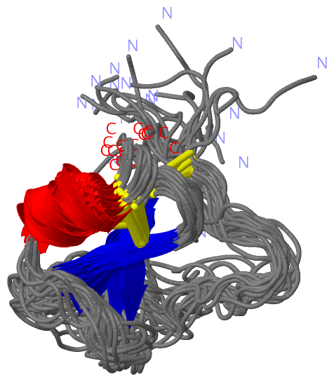
NMR Structure
|
||||||||||||||||
Cis Peptide Bonds (0, 0)| (no "Cis Peptide Bond" information available for 2LFL) |
SAPs(SNPs)/Variants (0, 0)| (no "SAP(SNP)/Variant" information available for 2LFL) |
PROSITE Motifs (0, 0)| (no "PROSITE Motif" information available for 2LFL) |
Exons (0, 0)| (no "Exon" information available for 2LFL) |
Sequences/Alignments
NMR StructureChain A from PDB Type:PROTEIN Length:57 aligned with Q1EG59_RHIAP | Q1EG59 from UniProtKB/TrEMBL Length:118 Alignment length:58 48 58 68 78 88 Q1EG59_RHIAP 39 GKRKEECTVPIGWSEPVKGLCKARFTRYYCMGNCCKVYEGCYTGGYSRMGECARNCPG 96 SCOP domains d2 lfla_ A: automated matches SCOP domains CATH domains ---------------------------------------------------------- CATH domains Pfam domains ---------------------------------------------------------- Pfam domains SAPs(SNPs) ---------------------------------------------------------- SAPs(SNPs) PROSITE ---------------------------------------------------------- PROSITE Transcript ---------------------------------------------------------- Transcript 2lfl A 19 GD-KEECTVPIGWSEPVKGLCKARFTRYYCMGNCCKVYEGCYTGGYSRMGECARNCPG 75 | | 27 37 47 57 67 | | 20 | 21
|
||||||||||||||||||||
SCOP Domains (1, 1)
NMR Structure
 |
CATH Domains (0, 0)| (no "CATH Domain" information available for 2LFL) |
Pfam Domains (0, 0)| (no "Pfam Domain" information available for 2LFL) |
Gene Ontology (0, 0)|
NMR Structure(hide GO term definitions)
(no "Gene Ontology" information available for 2LFL)
|
Interactive Views
|
||||||||||||||||||||||||||||||||||||||||||||||||||||||||||||||||||||||||||||||||||||||||||||||||||||||||||||||||||||
Still Images
|
||||||||||||||||
Databases
|
||||||||||||||||||||||||||||||||||||||||||||||||||||||||||||||||||||||||||||||||||||||||||||||||||||||||||||||||||||||||||||||||||||||||||||||||||||||||||||||||
Analysis Tools
|
|||||||||||||||||||||||||||||||||||||||||||||||||||||||||||||
Entries Sharing at Least One Protein Chain (UniProt ID)
Related Entries Specified in the PDB File
|
|