|
|
|
|
 Description
|
|
Compounds
|
||||||||||||||||||||||||||||||||||||||||||||
Chains, Units
Summary Information (see also Sequences/Alignments below) |
Ligands, Modified Residues, Ions (6, 24)
NMR Structure (6, 24)
|
Sites (6, 6)
NMR Structure (6, 6)
|
SS Bonds (0, 0)| (no "SS Bond" information available for 2L65) |
Cis Peptide Bonds (0, 0)| (no "Cis Peptide Bond" information available for 2L65) |
SAPs(SNPs)/Variants (0, 0)| (no "SAP(SNP)/Variant" information available for 2L65) |
PROSITE Motifs (0, 0)| (no "PROSITE Motif" information available for 2L65) |
Exons (0, 0)| (no "Exon" information available for 2L65) |
Sequences/Alignments
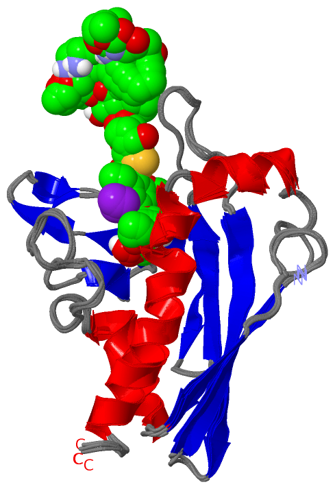
NMR StructureChain A from PDB Type:PROTEIN Length:155 aligned with Q8KNF0_MICEC | Q8KNF0 from UniProtKB/TrEMBL Length:181 Alignment length:155 36 46 56 66 76 86 96 106 116 126 136 146 156 166 176 Q8KNF0_MICEC 27 NYDPFVRHSVTVKADRKTAFKTFLEGFPEWWPNNFRTTKVGAPLGVDKKGGRWYEIDEQGEEHTFGLIRKVDEPDTLVIGWRLNGFGRIDPDNSSEFTVTFVADGQKKTRVDVEHTHFDRMGTKHAKRVRNGMDKGWPTILQSFQDKIDEEGAKK 181 SCOP domains d2l65a1 A:1-155 Calicheamicin gene cluster protein CalC SCOP domains CATH domains ----------------------------------------------------------------------------------------------------------------------------------------------------------- CATH domains Pfam domains ------------AHSA1-2l65A01 A:13-148 ------- Pfam domains SAPs(SNPs) ----------------------------------------------------------------------------------------------------------------------------------------------------------- SAPs(SNPs) PROSITE ----------------------------------------------------------------------------------------------------------------------------------------------------------- PROSITE Transcript ----------------------------------------------------------------------------------------------------------------------------------------------------------- Transcript 2l65 A 1 NYDPFVRHSVTVKADRKTAFKTFLEGFPEWWPNNFRTTKVGAPLGVDKKGGRWYEIDEQGEEHTFGLIRKVDEPDTLVIGWRLNGFGRIDPDNSSEFTVTFVADGQKKTRVDVEHTHFDRMGTKHAKRVRNGMDKGWPTILQSFQDKIDEEGAKK 155 10 20 30 40 50 60 70 80 90 100 110 120 130 140 150
|
||||||||||||||||||||
SCOP Domains (1, 1)
NMR Structure
 |
CATH Domains (0, 0)| (no "CATH Domain" information available for 2L65) |
Pfam Domains (1, 1)| NMR Structure |
Gene Ontology (0, 0)|
NMR Structure(hide GO term definitions)
(no "Gene Ontology" information available for 2L65)
|
Interactive Views
|
||||||||||||||||||||||||||||||||||||||||||||||||||||||||||||||||||||||||||||||||||||||||||||||||||||||||||||||||||||||||||||||||||||||||||||||||||||||||||||||||||||||||||||||||||||||||||||
Still Images
|
||||||||||||||||
Databases
|
||||||||||||||||||||||||||||||||||||||||||||||||||||||||||||||||||||||||||||||||||||||||||||||||||||||||||||||||||||||||||||||||||||||||||||||||||||||||||||||||
Analysis Tools
|
|||||||||||||||||||||||||||||||||||||||||||||||||||||||||||||
Entries Sharing at Least One Protein Chain (UniProt ID)
Related Entries Specified in the PDB File
|
|