|
|
|
|
 Description
|
|
Compounds
|
||||||||||||||||||||||||||||||||||||||||||||||||
Chains, Units
Summary Information (see also Sequences/Alignments below) |
Ligands, Modified Residues, Ions (0, 0)| (no "Ligand,Modified Residues,Ions" information available for 2L3W) |
Sites (0, 0)| (no "Site" information available for 2L3W) |
SS Bonds (0, 0)| (no "SS Bond" information available for 2L3W) |
Cis Peptide Bonds (0, 0)| (no "Cis Peptide Bond" information available for 2L3W) |
SAPs(SNPs)/Variants (0, 0)| (no "SAP(SNP)/Variant" information available for 2L3W) |
PROSITE Motifs (0, 0)| (no "PROSITE Motif" information available for 2L3W) |
Exons (0, 0)| (no "Exon" information available for 2L3W) |
Sequences/Alignments
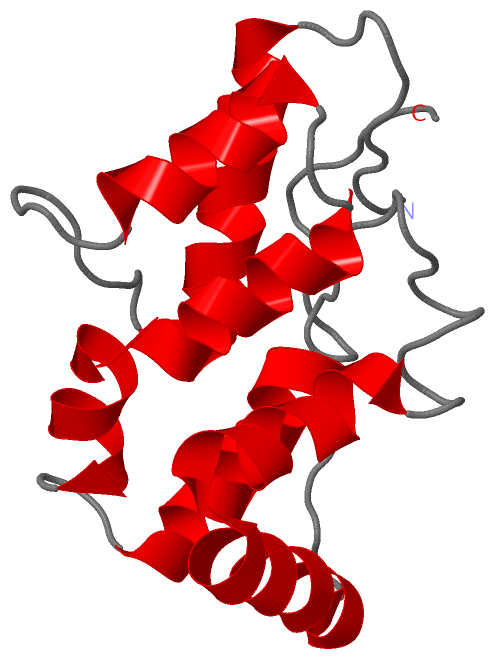
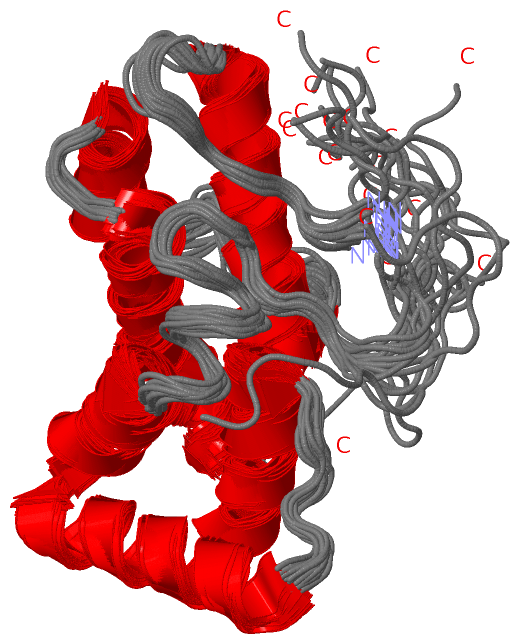
NMR StructureChain A from PDB Type:PROTEIN Length:143 aligned with Q31PE0_SYNE7 | Q31PE0 from UniProtKB/TrEMBL Length:273 Alignment length:143 28 38 48 58 68 78 88 98 108 118 128 138 148 158 Q31PE0_SYNE7 19 APLEWRAGASSDEINAIIRAVYRQVLGNDYVMSTERLTSAESLLRGGEISVRDFVRAVALSELYREKFFHNNAHNRFIELNFKHLLGRAPYDQAEVAAHAATYHSHGYDADINSYIDSAEYTESFGDNVVPYFRGFATIRAQK 161 SCOP domains ----------------------------------------------------------------------------------------------------------------------------------------------- SCOP domains CATH domains ----------------------------------------------------------------------------------------------------------------------------------------------- CATH domains Pfam domains ----PBS_linker_poly-2l3wA01 A:5-135 -------- Pfam domains SAPs(SNPs) ----------------------------------------------------------------------------------------------------------------------------------------------- SAPs(SNPs) PROSITE ----------------------------------------------------------------------------------------------------------------------------------------------- PROSITE Transcript ----------------------------------------------------------------------------------------------------------------------------------------------- Transcript 2l3w A 1 MPLEWRAGASSDEINAIIRAVYRQVLGNDYVMSTERLTSAESLLRGGEISVRDFVRAVALSELYREKFFHNNAHNRFIELNFKHLLGRAPYDQAEVAAHAATYHSHGYDADINSYIDSAEYTESFGDNVVPYFRGLEHHHHHH 143 10 20 30 40 50 60 70 80 90 100 110 120 130 140
|
||||||||||||||||||||
SCOP Domains (0, 0)| (no "SCOP Domain" information available for 2L3W) |
CATH Domains (0, 0)| (no "CATH Domain" information available for 2L3W) |
Pfam Domains (1, 1)
NMR Structure
|
Gene Ontology (2, 2)|
NMR Structure(hide GO term definitions) Chain A (Q31PE0_SYNE7 | Q31PE0)
|
||||||||||||||||||||||||
Interactive Views
|
||||||||||||||||||||||||||||||||||||||||||||||||||||||||||||||||||||||||||||||||||||||||||||||||||||||||||||||||||||
Still Images
|
||||||||||||||||
Databases
|
||||||||||||||||||||||||||||||||||||||||||||||||||||||||||||||||||||||||||||||||||||||||||||||||||||||||||||||||||||||||||||||||||||||||||||||||||||||||||||||||
Analysis Tools
|
|||||||||||||||||||||||||||||||||||||||||||||||||||||||||||||
Entries Sharing at Least One Protein Chain (UniProt ID)
Related Entries Specified in the PDB File
|
|