| molecular function |
|---|
| | GO:0003779 | | actin binding | | Interacting selectively and non-covalently with monomeric or multimeric forms of actin, including actin filaments. |
| | GO:0046872 | | metal ion binding | | Interacting selectively and non-covalently with any metal ion. |
| | GO:0016491 | | oxidoreductase activity | | Catalysis of an oxidation-reduction (redox) reaction, a reversible chemical reaction in which the oxidation state of an atom or atoms within a molecule is altered. One substrate acts as a hydrogen or electron donor and becomes oxidized, while the other acts as hydrogen or electron acceptor and becomes reduced. |
| | GO:0016671 | | oxidoreductase activity, acting on a sulfur group of donors, disulfide as acceptor | | Catalysis of an oxidation-reduction (redox) reaction in which a sulfur-containing group acts as a hydrogen or electron donor and reduces disulfide. |
| | GO:0033743 | | peptide-methionine (R)-S-oxide reductase activity | | Catalysis of the reaction: peptide-L-methionine + H(2)O + thioredoxin disulfide = peptide-L-methionine (R)-S-oxide + thioredoxin. Can act on oxidized methionine in peptide linkage with specificity for the R enantiomer. Thioredoxin disulfide is the oxidized form of thioredoxin. |
| | GO:0008270 | | zinc ion binding | | Interacting selectively and non-covalently with zinc (Zn) ions. |
| biological process |
|---|
| | GO:0030041 | | actin filament polymerization | | Assembly of actin filaments by the addition of actin monomers to a filament. |
| | GO:0055114 | | oxidation-reduction process | | A metabolic process that results in the removal or addition of one or more electrons to or from a substance, with or without the concomitant removal or addition of a proton or protons. |
| | GO:0030091 | | protein repair | | The process of restoring a protein to its original state after damage by such things as oxidation or spontaneous decomposition of residues. |
| | GO:0006979 | | response to oxidative stress | | Any process that results in a change in state or activity of a cell or an organism (in terms of movement, secretion, enzyme production, gene expression, etc.) as a result of oxidative stress, a state often resulting from exposure to high levels of reactive oxygen species, e.g. superoxide anions, hydrogen peroxide (H2O2), and hydroxyl radicals. |
| cellular component |
|---|
| | GO:0005739 | | mitochondrion | | A semiautonomous, self replicating organelle that occurs in varying numbers, shapes, and sizes in the cytoplasm of virtually all eukaryotic cells. It is notably the site of tissue respiration. |
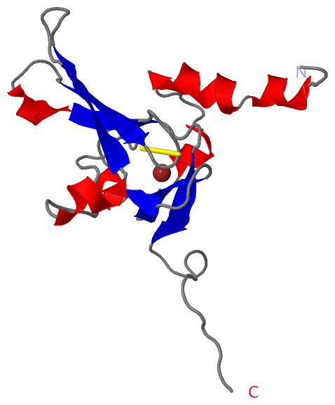
 Description
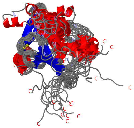