| molecular function |
|---|
| | GO:0019003 | | GDP binding | | Interacting selectively and non-covalently with GDP, guanosine 5'-diphosphate. |
| | GO:0005525 | | GTP binding | | Interacting selectively and non-covalently with GTP, guanosine triphosphate. |
| | GO:0003924 | | GTPase activity | | Catalysis of the reaction: GTP + H2O = GDP + phosphate. |
| | GO:0046872 | | metal ion binding | | Interacting selectively and non-covalently with any metal ion. |
| | GO:0000166 | | nucleotide binding | | Interacting selectively and non-covalently with a nucleotide, any compound consisting of a nucleoside that is esterified with (ortho)phosphate or an oligophosphate at any hydroxyl group on the ribose or deoxyribose. |
| | GO:0005515 | | protein binding | | Interacting selectively and non-covalently with any protein or protein complex (a complex of two or more proteins that may include other nonprotein molecules). |
| biological process |
|---|
| | GO:0032008 | | positive regulation of TOR signaling | | Any process that activates or increases the frequency, rate or extent of TOR signaling. |
| | GO:0048168 | | regulation of neuronal synaptic plasticity | | A process that modulates neuronal synaptic plasticity, the ability of neuronal synapses to change as circumstances require. They may alter function, such as increasing or decreasing their sensitivity, or they may increase or decrease in actual numbers. |
| | GO:0007165 | | signal transduction | | The cellular process in which a signal is conveyed to trigger a change in the activity or state of a cell. Signal transduction begins with reception of a signal (e.g. a ligand binding to a receptor or receptor activation by a stimulus such as light), or for signal transduction in the absence of ligand, signal-withdrawal or the activity of a constitutively active receptor. Signal transduction ends with regulation of a downstream cellular process, e.g. regulation of transcription or regulation of a metabolic process. Signal transduction covers signaling from receptors located on the surface of the cell and signaling via molecules located within the cell. For signaling between cells, signal transduction is restricted to events at and within the receiving cell. |
| | GO:0007264 | | small GTPase mediated signal transduction | | Any series of molecular signals in which a small monomeric GTPase relays one or more of the signals. |
| cellular component |
|---|
| | GO:0005794 | | Golgi apparatus | | A compound membranous cytoplasmic organelle of eukaryotic cells, consisting of flattened, ribosome-free vesicles arranged in a more or less regular stack. The Golgi apparatus differs from the endoplasmic reticulum in often having slightly thicker membranes, appearing in sections as a characteristic shallow semicircle so that the convex side (cis or entry face) abuts the endoplasmic reticulum, secretory vesicles emerging from the concave side (trans or exit face). In vertebrate cells there is usually one such organelle, while in invertebrates and plants, where they are known usually as dictyosomes, there may be several scattered in the cytoplasm. The Golgi apparatus processes proteins produced on the ribosomes of the rough endoplasmic reticulum; such processing includes modification of the core oligosaccharides of glycoproteins, and the sorting and packaging of proteins for transport to a variety of cellular locations. Three different regions of the Golgi are now recognized both in terms of structure and function: cis, in the vicinity of the cis face, trans, in the vicinity of the trans face, and medial, lying between the cis and trans regions. |
| | GO:0000139 | | Golgi membrane | | The lipid bilayer surrounding any of the compartments of the Golgi apparatus. |
| | GO:0005737 | | cytoplasm | | All of the contents of a cell excluding the plasma membrane and nucleus, but including other subcellular structures. |
| | GO:0005829 | | cytosol | | The part of the cytoplasm that does not contain organelles but which does contain other particulate matter, such as protein complexes. |
| | GO:0030425 | | dendrite | | A neuron projection that has a short, tapering, often branched, morphology, receives and integrates signals from other neurons or from sensory stimuli, and conducts a nerve impulse towards the axon or the cell body. In most neurons, the impulse is conveyed from dendrites to axon via the cell body, but in some types of unipolar neuron, the impulse does not travel via the cell body. |
| | GO:0012505 | | endomembrane system | | A collection of membranous structures involved in transport within the cell. The main components of the endomembrane system are endoplasmic reticulum, Golgi bodies, vesicles, cell membrane and nuclear envelope. Members of the endomembrane system pass materials through each other or though the use of vesicles. |
| | GO:0005783 | | endoplasmic reticulum | | The irregular network of unit membranes, visible only by electron microscopy, that occurs in the cytoplasm of many eukaryotic cells. The membranes form a complex meshwork of tubular channels, which are often expanded into slitlike cavities called cisternae. The ER takes two forms, rough (or granular), with ribosomes adhering to the outer surface, and smooth (with no ribosomes attached). |
| | GO:0005789 | | endoplasmic reticulum membrane | | The lipid bilayer surrounding the endoplasmic reticulum. |
| | GO:0016020 | | membrane | | A lipid bilayer along with all the proteins and protein complexes embedded in it an attached to it. |
| | GO:0043025 | | neuronal cell body | | The portion of a neuron that includes the nucleus, but excludes cell projections such as axons and dendrites. |
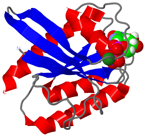
 Description
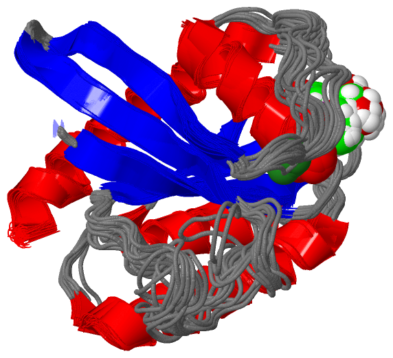