|
|
|
|
 Description
|
|
Compounds
|
||||||||||||||||||||||||||||||||||||||||||||||||
Chains, Units
Summary Information (see also Sequences/Alignments below) |
Ligands, Modified Residues, Ions (0, 0)| (no "Ligand,Modified Residues,Ions" information available for 2L06) |
Sites (0, 0)| (no "Site" information available for 2L06) |
SS Bonds (0, 0)| (no "SS Bond" information available for 2L06) |
Cis Peptide Bonds (0, 0)| (no "Cis Peptide Bond" information available for 2L06) |
SAPs(SNPs)/Variants (0, 0)| (no "SAP(SNP)/Variant" information available for 2L06) |
PROSITE Motifs (0, 0)| (no "PROSITE Motif" information available for 2L06) |
Exons (0, 0)| (no "Exon" information available for 2L06) |
Sequences/Alignments
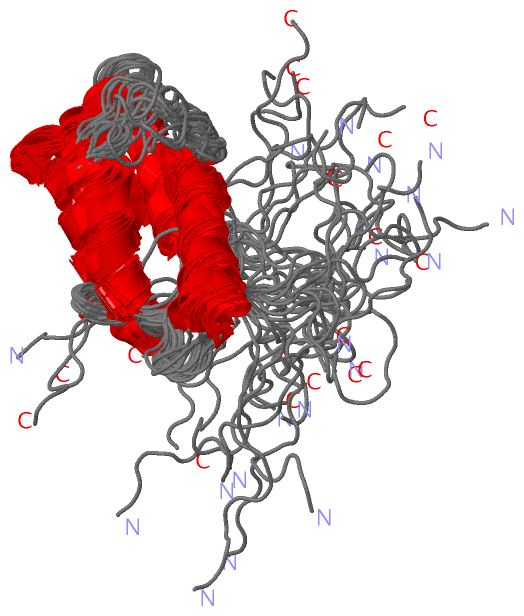
NMR StructureChain A from PDB Type:PROTEIN Length:155 aligned with APCE_SYNY3 | Q55544 from UniProtKB/Swiss-Prot Length:896 Alignment length:157 263 273 283 293 303 313 323 333 343 353 363 373 383 393 403 APCE_SYNY3 254 PQSYFNAAAKRQKYAMKPGLSALEKNAVIKAAYRQIFERDITKAYSQSISYLESQVRNGDISMKEFVRRLAKSPLYRKQFFEPFINSRALELAFRHILGRGPSSREEVQKYFSIVSSGGLPALVDALVDSQEYADYFGEETVPYLRGLGVEAQECRN 410 SCOP domains ------------------------------------------------------------------------------------------------------------------------------------------------------------- SCOP domains CATH domains ------------------------------------------------------------------------------------------------------------------------------------------------------------- CATH domains Pfam domains ---------------PBS_linker_poly-2l06A01 A:16-147 ---------- Pfam domains SAPs(SNPs) ------------------------------------------------------------------------------------------------------------------------------------------------------------- SAPs(SNPs) PROSITE ------------------------------------------------------------------------------------------------------------------------------------------------------------- PROSITE Transcript ------------------------------------------------------------------------------------------------------------------------------------------------------------- Transcript 2l06 A 1 PQSYFNAAAKRQKYAMKPGLSALEKNAVIKAAYRQIFERDITKAYSQSISYLESQVRNGDISMKEFVRRLAKSPLYRKQFFEPFINSRALELAFRHILGRGPSSREEVQKYFSIVSSGGLPALVDALVDSQEYADYFGEETVPYLRGL--EHHHHHH 155 10 20 30 40 50 60 70 80 90 100 110 120 130 140 | -| 148 | 149
|
||||||||||||||||||||
SCOP Domains (0, 0)| (no "SCOP Domain" information available for 2L06) |
CATH Domains (0, 0)| (no "CATH Domain" information available for 2L06) |
Pfam Domains (1, 1)
NMR Structure
|
Gene Ontology (9, 9)|
NMR Structure(hide GO term definitions) Chain A (APCE_SYNY3 | Q55544)
|
||||||||||||||||||||||||||||||||||||||||||||||||||||||||||||||||||||||||
Interactive Views
|
||||||||||||||||||||||||||||||||||||||||||||||||||||||||||||||||||||||||||||||||||||||||||||||||||||||||||||||||||||
Still Images
|
||||||||||||||||
Databases
|
||||||||||||||||||||||||||||||||||||||||||||||||||||||||||||||||||||||||||||||||||||||||||||||||||||||||||||||||||||||||||||||||||||||||||||||||||||||||||||||||
Analysis Tools
|
|||||||||||||||||||||||||||||||||||||||||||||||||||||||||||||
Entries Sharing at Least One Protein Chain (UniProt ID)
Related Entries Specified in the PDB File
|
|