Show PDB file:   
         Plain Text   HTML   (compressed file size)
QuickSearch:   
by PDB,NDB,UniProt,PROSITE Code or Search Term(s)  
(-)NMR Structure - model 1
(-)NMR Structure - all models
collapse expand < >
Image NMR Structure - model 1
NMR Structure - model 1  (Jmol Viewer)
Image NMR Structure - all models
NMR Structure - all models  (Jmol Viewer)

(-) Description

Title :  SOLUTION STRUCTURE OF UBX DOMAIN OF HUMAN UBXD2 PROTEIN
 
Authors :  Q. Wu, H. Huang, J. Zhang, Q. Hu, J. Wu, Y. Shi
Date :  06 May 10  (Deposition) - 18 May 11  (Release) - 18 May 11  (Revision)
Method :  SOLUTION NMR
Resolution :  NOT APPLICABLE
Chains :  NMR Structure  :  A  (20x)
NMR Structure *:  A  (1x)
Keywords :  Ubxd2, Ubx, Vcp, P97, Protein Binding (Keyword Search: [Gene Ontology, PubMed, Web (Google))
 
Reference :  Q. Wu, H. Huang, J. Zhang, Q. Hu, J. Wu, Y. Shi
Solution Strcture Of Ubx Domain Of Human Ubxd2 Protein
To Be Published
PubMed: search

(-) Compounds

Molecule 1 - UBX DOMAIN-CONTAINING PROTEIN 4
    ChainsA
    EngineeredYES
    Expression SystemESCHERICHIA COLI
    Expression System Taxid562
    Expression System VectorP28
    Expression System Vector TypeVECTOR
    FragmentUBX DOMAIN, UNP RESIDUES 317-397
    GeneUBXN4, KIAA0242, UBXD2, UBXDC1
    Organism CommonHUMAN
    Organism ScientificHOMO SAPIENS
    Organism Taxid9606
    SynonymUBX DOMAIN-CONTAINING PROTEIN 2, ERASIN

 Structural Features

(-) Chains, Units

  1
NMR Structure (20x)A
NMR Structure * (1x)A

Summary Information (see also Sequences/Alignments below)

(-) Ligands, Modified Residues, Ions  (0, 0)

(no "Ligand,Modified Residues,Ions" information available for 2KXJ)

(-) Sites  (0, 0)

(no "Site" information available for 2KXJ)

(-) SS Bonds  (0, 0)

(no "SS Bond" information available for 2KXJ)

(-) Cis Peptide Bonds  (2, 40)
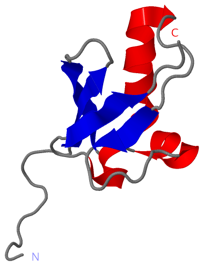
NMR Structure
No.ModelResidues
11, 2, 3, 4, 5, 6, 7, 8, 9, 10, 11, 12, 13, 14, 15, 16, 17, 18, 19, 20Phe A:366 -Pro A:367
21, 2, 3, 4, 5, 6, 7, 8, 9, 10, 11, 12, 13, 14, 15, 16, 17, 18, 19, 20Ala A:386 -Pro A:387

 Sequence-Structure Mapping

(-) SAPs(SNPs)/Variants  (0, 0)

(no "SAP(SNP)/Variant" information available for 2KXJ)

(-) PROSITE Motifs  (1, 1)

NMR Structure (1, 1)
 PROSITEUniProtKBPDB
No.IDACDescriptionIDLocationCountLocation
1UBXPS50033 UBX domain profile.UBXN4_HUMAN315-393  1A:317-393
NMR Structure * (1, 1)
 PROSITEUniProtKBPDB
No.IDACDescriptionIDLocationCountLocation
1UBXPS50033 UBX domain profile.UBXN4_HUMAN315-393  1A:317-393

(-) Exons   (0, 0)

(no "Exon" information available for 2KXJ)

(-) Sequences/Alignments

NMR Structure
   Reformat: Number of residues per line =  ('0' or empty: single-line sequence representation)
  Number of residues per labelling interval =   
  UniProt sequence: complete  aligned part    
   Show mapping: SCOP domains CATH domains Pfam domains Secondary structure (by author)
SAPs(SNPs) PROSITE motifs Exons
(details for a mapped element are shown in a popup box when the mouse pointer rests over it)
Chain A from PDB  Type:PROTEIN  Length:90
 aligned with UBXN4_HUMAN | Q92575 from UniProtKB/Swiss-Prot  Length:508

    Alignment length:90
                                   317       327       337       347       357       367       377       387       397
          UBXN4_HUMAN   308 KRESYARERSTVARIQFRLPDGSSFTNQFPSDAPLEEARQFAAQTVGNTYGNFSLATMFPRREFTKEDYKKKLLDLELAPSASVVLLPAG 397
               SCOP domains ------------------------------------------------------------------------------------------ SCOP domains
               CATH domains ------------------------------------------------------------------------------------------ CATH domains
               Pfam domains ---------UBX-2kxjA01 A:317-395                                                          -- Pfam domains
         Sec.struct. author ...........eeeeeee.....eeeeeee....hhhhhhhhhhhhh.......eee.......hhhhhhh.hhhhh....eeeee.... Sec.struct. author
                 SAPs(SNPs) ------------------------------------------------------------------------------------------ SAPs(SNPs)
                    PROSITE -------UBX  PDB: A:317-393 UniProt: 315-393                                           ---- PROSITE
                 Transcript ------------------------------------------------------------------------------------------ Transcript
                 2kxj A 308 MGHHHHHHMSTVARIQFRLPDGSSFTNQFPSDAPLEEARQFAAQTVGNTYGNFSLATMFPRREFTKEDYKKKLLDLELAPSASVVLLPAG 397
                                   317       327       337       347       357       367       377       387       397

   Legend:   → Mismatch (orange background)
  - → Gap (green background, '-', border residues have a numbering label)
    → Modified Residue (blue background, lower-case, 'x' indicates undefined single-letter code, labelled with number + name)
  x → Chemical Group (purple background, 'x', labelled with number + name, e.g. ACE or NH2)
  extra numbering lines below/above indicate numbering irregularities and modified residue names etc., number ends below/above '|'

 Classification and Annotation

(-) SCOP Domains  (0, 0)

(no "SCOP Domain" information available for 2KXJ)

(-) CATH Domains  (0, 0)

(no "CATH Domain" information available for 2KXJ)

(-) Pfam Domains  (1, 1)

NMR Structure
(-)
Clan: Ubiquitin (279)

(-) Gene Ontology  (9, 9)

NMR Structure(hide GO term definitions)
Chain A   (UBXN4_HUMAN | Q92575)
molecular function
    GO:0005515    protein binding    Interacting selectively and non-covalently with any protein or protein complex (a complex of two or more proteins that may include other nonprotein molecules).
biological process
    GO:0006986    response to unfolded protein    Any process that results in a change in state or activity of a cell or an organism (in terms of movement, secretion, enzyme production, gene expression, etc.) as a result of an unfolded protein stimulus.
    GO:0030433    ubiquitin-dependent ERAD pathway    The series of steps necessary to target endoplasmic reticulum (ER)-resident proteins for degradation by the cytoplasmic proteasome. Begins with recognition of the ER-resident protein, includes retrotranslocation (dislocation) of the protein from the ER to the cytosol, protein ubiquitination necessary for correct substrate transfer, transport of the protein to the proteasome, and ends with degradation of the protein by the cytoplasmic proteasome.
cellular component
    GO:0005737    cytoplasm    All of the contents of a cell excluding the plasma membrane and nucleus, but including other subcellular structures.
    GO:0005783    endoplasmic reticulum    The irregular network of unit membranes, visible only by electron microscopy, that occurs in the cytoplasm of many eukaryotic cells. The membranes form a complex meshwork of tubular channels, which are often expanded into slitlike cavities called cisternae. The ER takes two forms, rough (or granular), with ribosomes adhering to the outer surface, and smooth (with no ribosomes attached).
    GO:0005789    endoplasmic reticulum membrane    The lipid bilayer surrounding the endoplasmic reticulum.
    GO:0016020    membrane    A lipid bilayer along with all the proteins and protein complexes embedded in it an attached to it.
    GO:0005635    nuclear envelope    The double lipid bilayer enclosing the nucleus and separating its contents from the rest of the cytoplasm; includes the intermembrane space, a gap of width 20-40 nm (also called the perinuclear space).
    GO:0005634    nucleus    A membrane-bounded organelle of eukaryotic cells in which chromosomes are housed and replicated. In most cells, the nucleus contains all of the cell's chromosomes except the organellar chromosomes, and is the site of RNA synthesis and processing. In some species, or in specialized cell types, RNA metabolism or DNA replication may be absent.

 Visualization

(-) Interactive Views

NMR Structure
  Complete Structure
    Jena3D(integrated viewing of ligand, site, SAP, PROSITE, SCOP information)
    WebMol | AstexViewer[tm]@PDBe
(Java Applets, require no local installation except for Java; loading may be slow)
    STRAP
(Java WebStart application, automatic local installation, requires Java; full application with system access!)
    RasMol
(require local installation)
    Molscript (VRML)
(requires installation of a VRML viewer; select preferred view via VRML and generate a mono or stereo PDF format file)
 
  Ligands, Modified Residues, Ions
(no "Ligands, Modified Residues, Ions" information available for 2kxj)
 
  Sites
(no "Sites" information available for 2kxj)
 
  Cis Peptide Bonds
    Ala A:386 - Pro A:387   [ RasMol ]  
    Phe A:366 - Pro A:367   [ RasMol ]  
 

(-) Still Images

Jmol
  protein: cartoon or spacefill or dots and stick; nucleic acid: cartoon and stick; ligands: spacefill; active site: stick
Molscript
  protein, nucleic acid: cartoon; ligands: spacefill; active site: ball and stick

 Databases and Analysis Tools

(-) Databases

Access by PDB/NDB ID
  2kxj
    Family and Domain InformationProDom | SYSTERS
    General Structural InformationGlycoscienceDB | MMDB | NDB | OCA | PDB | PDBe | PDBj | PDBsum | PDBWiki | PQS | PROTEOPEDIA
    Orientation in MembranesOPM
    Protein SurfaceSURFACE
    Secondary StructureDSSP (structure derived) | HSSP (homology derived)
    Structural GenomicsGeneCensus
    Structural NeighboursCE | VAST
    Structure ClassificationCATH | Dali | SCOP
    Validation and Original DataBMRB Data View | BMRB Restraints Grid | EDS | PROCHECK | RECOORD | WHAT_CHECK
 
Access by UniProt ID/Accession number
  UBXN4_HUMAN | Q92575
    Comparative Protein Structure ModelsModBase
    Genomic InformationEnsembl
    Protein-protein InteractionDIP
    Sequence, Family and Domain InformationInterPro | Pfam | SMART | UniProtKB/SwissProt
 
Access by Enzyme Classificator   (EC Number)
  (no 'Enzyme Classificator' available)
    General Enzyme InformationBRENDA | EC-PDB | Enzyme | IntEnz
    PathwayKEGG | MetaCyc
 
Access by Disease Identifier   (MIM ID)
  (no 'MIM ID' available)
    Disease InformationOMIM
 
Access by GenAge ID
  (no 'GenAge ID' available)
    Age Related InformationGenAge

(-) Analysis Tools

Access by PDB/NDB ID
    Domain InformationXDom
    Interatomic Contacts of Structural UnitsCSU
    Ligand-protein ContactsLPC
    Protein CavitiescastP
    Sequence and Secondary StructurePDBCartoon
    Structure AlignmentSTRAP(Java WebStart application, automatic local installation, requires Java; full application with system access!)
    Structure and Sequence BrowserSTING
 
Access by UniProt ID/Accession number
  UBXN4_HUMAN | Q92575
    Protein Disorder PredictionDisEMBL | FoldIndex | GLOBPLOT (for more information see DisProt)

 Related Entries

(-) Entries Sharing at Least One Protein Chain (UniProt ID)

(no "Entries Sharing at Least One Protein Chain" available for 2KXJ)

(-) Related Entries Specified in the PDB File

(no "Related Entries Specified in the PDB File" available for 2KXJ)