Show PDB file:   
         Plain Text   HTML   (compressed file size)
QuickSearch:   
by PDB,NDB,UniProt,PROSITE Code or Search Term(s)  
(-)NMR Structure - model 1
(-)NMR Structure - all models
collapse expand < >
Image NMR Structure - model 1
NMR Structure - model 1  (Jmol Viewer)
Image NMR Structure - all models
NMR Structure - all models  (Jmol Viewer)

(-) Description

Title :  NMR STRUCTURE OF MOUSE PAR3-PDZ3 IN COMPLEX WITH VE-CADHERIN C-TERMINUS
 
Authors :  B. F. Volkman, R. C. Tyler, F. C. Peterson
Date :  22 Sep 09  (Deposition) - 09 Feb 10  (Release) - 23 Feb 10  (Revision)
Method :  SOLUTION NMR
Resolution :  NOT APPLICABLE
Chains :  NMR Structure  :  A,B  (20x)
Keywords :  Par3, Pdz Domain, Ve Cadherin, Alternative Splicing, Cell Cycle, Cell Division, Cell Junction, Coiled Coil, Cytoplasm, Cytoskeleton, Membrane, Phosphoprotein, Tight Junction, Calcium, Cell Adhesion, Cell Membrane, Cleavage On Pair Of Basic Residues, Glycoprotein, Transmembrane, Protein Binding (Keyword Search: [Gene Ontology, PubMed, Web (Google))
 
Reference :  R. C. Tyler, F. C. Peterson, B. F. Volkman
Distal Interactions Within The Par3-Ve-Cadherin Complex.
Biochemistry V. 49 951 2010
PubMed-ID: 20047332  |  Reference-DOI: 10.1021/BI9017335

(-) Compounds

Molecule 1 - PARTITIONING DEFECTIVE 3 HOMOLOG
    ChainsA
    EngineeredYES
    Expression SystemESCHERICHIA COLI
    Expression System StrainSG130099[PREP4]
    Expression System Taxid562
    Expression System VectorPQE30-8HT
    FragmentUNP RESIDUES 581-689, PDZ 3 DOMAIN
    GenePAR3, PARD3
    Organism CommonMOUSE
    Organism ScientificMUS MUSCULUS
    Organism Taxid10090
    SynonymPARD-3, PAR-3, ATYPICAL PKC ISOTYPE-SPECIFIC- INTERACTING PROTEIN, ASIP, EPHRIN-INTERACTING PROTEIN, PHIP
 
Molecule 2 - CADHERIN-5
    ChainsB
    EngineeredYES
    Expression SystemESCHERICHIA COLI
    Expression System StrainSG130099[PREP4]
    Expression System Taxid562
    Expression System VectorPQE30-SMT
    FragmentUNP RESIDUES 769-784
    GeneCDH5, VE-CADHERIN
    Organism CommonMOUSE
    Organism ScientificMUS MUSCULUS
    Organism Taxid10090
    SynonymVASCULAR ENDOTHELIAL CADHERIN, VE-CADHERIN

 Structural Features

(-) Chains, Units

  
NMR Structure (20x)

Summary Information (see also Sequences/Alignments below)

(-) Ligands, Modified Residues, Ions  (0, 0)

(no "Ligand,Modified Residues,Ions" information available for 2KOH)

(-) Sites  (0, 0)

(no "Site" information available for 2KOH)

(-) SS Bonds  (0, 0)

(no "SS Bond" information available for 2KOH)

(-) Cis Peptide Bonds  (0, 0)

(no "Cis Peptide Bond" information available for 2KOH)

 Sequence-Structure Mapping

(-) SAPs(SNPs)/Variants  (0, 0)

(no "SAP(SNP)/Variant" information available for 2KOH)

(-) PROSITE Motifs  (1, 1)

NMR Structure (1, 1)
 PROSITEUniProtKBPDB
No.IDACDescriptionIDLocationCountLocation
1PDZPS50106 PDZ domain profile.PARD3_MOUSE271-347
461-546
590-665
  1-
-
A:590-665

(-) Exons   (0, 0)

(no "Exon" information available for 2KOH)

(-) Sequences/Alignments

NMR Structure
   Reformat: Number of residues per line =  ('0' or empty: single-line sequence representation)
  Number of residues per labelling interval =   
  UniProt sequence: complete  aligned part    
   Show mapping: SCOP domains CATH domains Pfam domains Secondary structure (by author)
SAPs(SNPs) PROSITE motifs Exons
(details for a mapped element are shown in a popup box when the mouse pointer rests over it)
Chain A from PDB  Type:PROTEIN  Length:111
 aligned with PARD3_MOUSE | Q99NH2 from UniProtKB/Swiss-Prot  Length:1333

    Alignment length:111
                                   588       598       608       618       628       638       648       658       668       678       688 
          PARD3_MOUSE   579 TPDGTREFLTFEVPLNDSGSAGLGVSVKGNRSKENHADLGIFVKSIINGGAASKDGRLRVNDQLIAVNGESLLGKANQEAMETLRRSMSTEGNKRGMIQLIVARRISRCNE 689
               SCOP domains d2koha_ A: automated matches                                                                                    SCOP domains
               CATH domains 2kohA00 A:579-689  [code=2.30.42.10, no name defined]                                                           CATH domains
               Pfam domains -----------PDZ-2kohA01 A:590-680                                                                      --------- Pfam domains
         Sec.struct. author .....eeeeeeeee..........eeeeeeee....eeeeeeeeee...hhhhhh.......eeeee..ee....hhhhhhhhhhhhh........eeeeeeeee...... Sec.struct. author
                 SAPs(SNPs) --------------------------------------------------------------------------------------------------------------- SAPs(SNPs)
                    PROSITE -----------PDZ  PDB: A:590-665 UniProt: 590-665                                        ------------------------ PROSITE
                 Transcript --------------------------------------------------------------------------------------------------------------- Transcript
                 2koh A 579 GSDGTREFLTFEVPLNDSGSAGLGVSVKGNRSKENHADLGIFVKSIINGGAASKDGRLRVNDQLIAVNGESLLGKANQEAMETLRRSMSTEGNKRGMIQLIVARRISRCNE 689
                                   588       598       608       618       628       638       648       658       668       678       688 

Chain B from PDB  Type:PROTEIN  Length:16
 aligned with CADH5_MOUSE | P55284 from UniProtKB/Swiss-Prot  Length:784

    Alignment length:16
                                   778      
          CADH5_MOUSE   769 MLAELYGSDPQEELII 784
               SCOP domains ---------------- SCOP domains
               CATH domains ---------------- CATH domains
               Pfam domains ---------------- Pfam domains
         Sec.struct. author ............eee. Sec.struct. author
                 SAPs(SNPs) ---------------- SAPs(SNPs)
                    PROSITE ---------------- PROSITE
                 Transcript ---------------- Transcript
                 2koh B 769 MLAELYGSDPQEELII 784
                                   778      

   Legend:   → Mismatch (orange background)
  - → Gap (green background, '-', border residues have a numbering label)
    → Modified Residue (blue background, lower-case, 'x' indicates undefined single-letter code, labelled with number + name)
  x → Chemical Group (purple background, 'x', labelled with number + name, e.g. ACE or NH2)
  extra numbering lines below/above indicate numbering irregularities and modified residue names etc., number ends below/above '|'

 Classification and Annotation

(-) SCOP Domains  (1, 1)

NMR Structure

(-) CATH Domains  (1, 1)

NMR Structure
(-)
Class: Mainly Beta (13760)

(-) Pfam Domains  (1, 1)

NMR Structure
(-)
Clan: PDZ-like (184)
(-)
Family: PDZ (172)

(-) Gene Ontology  (58, 66)

NMR Structure(hide GO term definitions)
Chain A   (PARD3_MOUSE | Q99NH2)
molecular function
    GO:0008289    lipid binding    Interacting selectively and non-covalently with a lipid.
    GO:0005547    phosphatidylinositol-3,4,5-trisphosphate binding    Interacting selectively and non-covalently with phosphatidylinositol-3,4,5-trisphosphate, a derivative of phosphatidylinositol in which the inositol ring is phosphorylated at the 3', 4' and 5' positions.
    GO:0032266    phosphatidylinositol-3-phosphate binding    Interacting selectively and non-covalently with phosphatidylinositol-3-phosphate, a derivative of phosphatidylinositol in which the inositol ring is phosphorylated at the 3' position.
    GO:0005546    phosphatidylinositol-4,5-bisphosphate binding    Interacting selectively and non-covalently with phosphatidylinositol-4,5-bisphosphate, a derivative of phosphatidylinositol in which the inositol ring is phosphorylated at the 4' and 5' positions.
    GO:0005515    protein binding    Interacting selectively and non-covalently with any protein or protein complex (a complex of two or more proteins that may include other nonprotein molecules).
    GO:0005080    protein kinase C binding    Interacting selectively and non-covalently with protein kinase C.
biological process
    GO:0003383    apical constriction    The actin-mediated process that results in the contraction of the apical end of a polarized columnar epithelial cell.
    GO:0070830    bicellular tight junction assembly    The aggregation, arrangement and bonding together of a set of components to form a tight junction, an occluding cell-cell junction that is composed of a branching network of sealing strands that completely encircles the apical end of each cell in an epithelial sheet.
    GO:0007049    cell cycle    The progression of biochemical and morphological phases and events that occur in a cell during successive cell replication or nuclear replication events. Canonically, the cell cycle comprises the replication and segregation of genetic material followed by the division of the cell, but in endocycles or syncytial cells nuclear replication or nuclear division may not be followed by cell division.
    GO:0030154    cell differentiation    The process in which relatively unspecialized cells, e.g. embryonic or regenerative cells, acquire specialized structural and/or functional features that characterize the cells, tissues, or organs of the mature organism or some other relatively stable phase of the organism's life history. Differentiation includes the processes involved in commitment of a cell to a specific fate and its subsequent development to the mature state.
    GO:0051301    cell division    The process resulting in division and partitioning of components of a cell to form more cells; may or may not be accompanied by the physical separation of a cell into distinct, individually membrane-bounded daughter cells.
    GO:0051642    centrosome localization    Any process in which a centrosome is transported to, and/or maintained in, a specific location within the cell.
    GO:0090162    establishment of epithelial cell polarity    The specification and formation of anisotropic intracellular organization of an epithelial cell.
    GO:0007163    establishment or maintenance of cell polarity    Any cellular process that results in the specification, formation or maintenance of anisotropic intracellular organization or cell growth patterns.
    GO:0000226    microtubule cytoskeleton organization    A process that is carried out at the cellular level which results in the assembly, arrangement of constituent parts, or disassembly of cytoskeletal structures comprising microtubules and their associated proteins.
    GO:0022011    myelination in peripheral nervous system    The process in which neuronal axons and dendrites become coated with a segmented lipid-rich sheath (myelin) to enable faster and more energetically efficient conduction of electrical impulses. The sheath is formed by the cell membranes of Schwann cells in the peripheral nervous system. Adjacent myelin segments are separated by a non-myelinated stretch of axon called a node of Ranvier.
    GO:0010801    negative regulation of peptidyl-threonine phosphorylation    Any process that decreases the frequency, rate or extent of peptidyl-threonine phosphorylation. Peptidyl-threonine phosphorylation is the phosphorylation of peptidyl-threonine to form peptidyl-O-phospho-L-threonine.
    GO:0031643    positive regulation of myelination    Any process that activates or increases the frequency, rate or extent of the formation of a myelin sheath around nerve axons.
    GO:0002092    positive regulation of receptor internalization    Any process that activates or increases the frequency, rate or extent of receptor internalization.
    GO:0006612    protein targeting to membrane    The process of directing proteins towards a membrane, usually using signals contained within the protein.
    GO:0032970    regulation of actin filament-based process    Any process that modulates the frequency, rate or extent of any cellular process that depends upon or alters the actin cytoskeleton.
    GO:0016337    single organismal cell-cell adhesion    The attachment of one cell to another cell via adhesion molecules, where both cells are part of the same organism.
    GO:0044319    wound healing, spreading of cells    The migration of a cell along or through a wound gap that contributes to the reestablishment of a continuous surface.
cellular component
    GO:0043220    Schmidt-Lanterman incisure    Regions within compact myelin in which the cytoplasmic faces of the enveloping myelin sheath are not tightly juxtaposed, and include cytoplasm from the cell responsible for making the myelin. Schmidt-Lanterman incisures occur in the compact myelin internode, while lateral loops are analogous structures found in the paranodal region adjacent to the nodes of Ranvier.
    GO:0005912    adherens junction    A cell junction at which anchoring proteins (cadherins or integrins) extend through the plasma membrane and are attached to actin filaments.
    GO:0045177    apical part of cell    The region of a polarized cell that forms a tip or is distal to a base. For example, in a polarized epithelial cell, the apical region has an exposed surface and lies opposite to the basal lamina that separates the epithelium from other tissue.
    GO:0005923    bicellular tight junction    An occluding cell-cell junction that is composed of a branching network of sealing strands that completely encircles the apical end of each cell in an epithelial sheet; the outer leaflets of the two interacting plasma membranes are seen to be tightly apposed where sealing strands are present. Each sealing strand is composed of a long row of transmembrane adhesion proteins embedded in each of the two interacting plasma membranes.
    GO:0005938    cell cortex    The region of a cell that lies just beneath the plasma membrane and often, but not always, contains a network of actin filaments and associated proteins.
    GO:0030054    cell junction    A cellular component that forms a specialized region of connection between two or more cells or between a cell and the extracellular matrix. At a cell junction, anchoring proteins extend through the plasma membrane to link cytoskeletal proteins in one cell to cytoskeletal proteins in neighboring cells or to proteins in the extracellular matrix.
    GO:0005913    cell-cell adherens junction    An adherens junction which connects a cell to another cell.
    GO:0005911    cell-cell junction    A cell junction that forms a connection between two or more cells in a multicellular organism; excludes direct cytoplasmic junctions such as ring canals.
    GO:0005737    cytoplasm    All of the contents of a cell excluding the plasma membrane and nucleus, but including other subcellular structures.
    GO:0005856    cytoskeleton    Any of the various filamentous elements that form the internal framework of cells, and typically remain after treatment of the cells with mild detergent to remove membrane constituents and soluble components of the cytoplasm. The term embraces intermediate filaments, microfilaments, microtubules, the microtrabecular lattice, and other structures characterized by a polymeric filamentous nature and long-range order within the cell. The various elements of the cytoskeleton not only serve in the maintenance of cellular shape but also have roles in other cellular functions, including cellular movement, cell division, endocytosis, and movement of organelles.
    GO:0012505    endomembrane system    A collection of membranous structures involved in transport within the cell. The main components of the endomembrane system are endoplasmic reticulum, Golgi bodies, vesicles, cell membrane and nuclear envelope. Members of the endomembrane system pass materials through each other or though the use of vesicles.
    GO:0033269    internode region of axon    An axon part that is located between the nodes of Ranvier and surrounded by compact myelin sheath.
    GO:0043219    lateral loop    Non-compact myelin located adjacent to the nodes of Ranvier in a myelin segment. These non-compact regions include cytoplasm from the cell responsible for synthesizing the myelin. Lateral loops are found in the paranodal region adjacent to the nodes of Ranvier, while Schmidt-Lantermann clefts are analogous structures found within the compact myelin internode.
    GO:0016020    membrane    A lipid bilayer along with all the proteins and protein complexes embedded in it an attached to it.
    GO:0005886    plasma membrane    The membrane surrounding a cell that separates the cell from its external environment. It consists of a phospholipid bilayer and associated proteins.
    GO:0043234    protein complex    A stable macromolecular complex composed (only) of two or more polypeptide subunits along with any covalently attached molecules (such as lipid anchors or oligosaccharide) or non-protein prosthetic groups (such as nucleotides or metal ions). Prosthetic group in this context refers to a tightly bound cofactor. The component polypeptide subunits may be identical.
    GO:0005819    spindle    The array of microtubules and associated molecules that forms between opposite poles of a eukaryotic cell during mitosis or meiosis and serves to move the duplicated chromosomes apart.
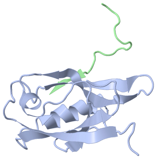
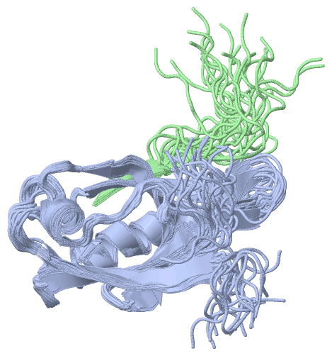
Chain B   (CADH5_MOUSE | P55284)
molecular function
    GO:0008013    beta-catenin binding    Interacting selectively and non-covalently with the beta subunit of the catenin complex.
    GO:0005509    calcium ion binding    Interacting selectively and non-covalently with calcium ions (Ca2+).
    GO:0044325    ion channel binding    Interacting selectively and non-covalently with one or more specific sites on an ion channel, a protein complex that spans a membrane and forms a water-filled channel across the phospholipid bilayer allowing selective ion transport down its electrochemical gradient.
    GO:0046872    metal ion binding    Interacting selectively and non-covalently with any metal ion.
    GO:0005515    protein binding    Interacting selectively and non-covalently with any protein or protein complex (a complex of two or more proteins that may include other nonprotein molecules).
    GO:0019903    protein phosphatase binding    Interacting selectively and non-covalently with any protein phosphatase.
    GO:0005102    receptor binding    Interacting selectively and non-covalently with one or more specific sites on a receptor molecule, a macromolecule that undergoes combination with a hormone, neurotransmitter, drug or intracellular messenger to initiate a change in cell function.
biological process
    GO:0001955    blood vessel maturation    A developmental process, independent of morphogenetic (shape) change, that is required for a blood vessel to attain its fully functional state.
    GO:0007155    cell adhesion    The attachment of a cell, either to another cell or to an underlying substrate such as the extracellular matrix, via cell adhesion molecules.
    GO:0007043    cell-cell junction assembly    The aggregation, arrangement and bonding together of a set of components to form a junction between cells.
    GO:0007156    homophilic cell adhesion via plasma membrane adhesion molecules    The attachment of a plasma membrane adhesion molecule in one cell to an identical molecule in an adjacent cell.
    GO:0008285    negative regulation of cell proliferation    Any process that stops, prevents or reduces the rate or extent of cell proliferation.
    GO:1903142    positive regulation of establishment of endothelial barrier    Any process that activates or increases the frequency, rate or extent of establishment of endothelial barrier.
    GO:2000114    regulation of establishment of cell polarity    Any process that modulates the frequency, rate or extent of establishment of cell polarity.
    GO:0016337    single organismal cell-cell adhesion    The attachment of one cell to another cell via adhesion molecules, where both cells are part of the same organism.
    GO:0007179    transforming growth factor beta receptor signaling pathway    A series of molecular signals initiated by the binding of an extracellular ligand to a transforming growth factor beta receptor on the surface of a target cell, and ending with regulation of a downstream cellular process, e.g. transcription.
cellular component
    GO:0005912    adherens junction    A cell junction at which anchoring proteins (cadherins or integrins) extend through the plasma membrane and are attached to actin filaments.
    GO:0005923    bicellular tight junction    An occluding cell-cell junction that is composed of a branching network of sealing strands that completely encircles the apical end of each cell in an epithelial sheet; the outer leaflets of the two interacting plasma membranes are seen to be tightly apposed where sealing strands are present. Each sealing strand is composed of a long row of transmembrane adhesion proteins embedded in each of the two interacting plasma membranes.
    GO:0030054    cell junction    A cellular component that forms a specialized region of connection between two or more cells or between a cell and the extracellular matrix. At a cell junction, anchoring proteins extend through the plasma membrane to link cytoskeletal proteins in one cell to cytoskeletal proteins in neighboring cells or to proteins in the extracellular matrix.
    GO:0071944    cell periphery    The part of a cell encompassing the cell cortex, the plasma membrane, and any external encapsulating structures.
    GO:0009986    cell surface    The external part of the cell wall and/or plasma membrane.
    GO:0005911    cell-cell junction    A cell junction that forms a connection between two or more cells in a multicellular organism; excludes direct cytoplasmic junctions such as ring canals.
    GO:0009897    external side of plasma membrane    The leaflet of the plasma membrane that faces away from the cytoplasm and any proteins embedded or anchored in it or attached to its surface.
    GO:0016021    integral component of membrane    The component of a membrane consisting of the gene products and protein complexes having at least some part of their peptide sequence embedded in the hydrophobic region of the membrane.
    GO:0016020    membrane    A lipid bilayer along with all the proteins and protein complexes embedded in it an attached to it.
    GO:0005886    plasma membrane    The membrane surrounding a cell that separates the cell from its external environment. It consists of a phospholipid bilayer and associated proteins.

 Visualization

(-) Interactive Views

NMR Structure
  Complete Structure
    Jena3D(integrated viewing of ligand, site, SAP, PROSITE, SCOP information)
    WebMol | AstexViewer[tm]@PDBe
(Java Applets, require no local installation except for Java; loading may be slow)
    STRAP
(Java WebStart application, automatic local installation, requires Java; full application with system access!)
    RasMol
(require local installation)
    Molscript (VRML)
(requires installation of a VRML viewer; select preferred view via VRML and generate a mono or stereo PDF format file)
 
  Ligands, Modified Residues, Ions
(no "Ligands, Modified Residues, Ions" information available for 2koh)
 
  Sites
(no "Sites" information available for 2koh)
 
  Cis Peptide Bonds
(no "Cis Peptide Bonds" information available for 2koh)
 

(-) Still Images

Jmol
  protein: cartoon or spacefill or dots and stick; nucleic acid: cartoon and stick; ligands: spacefill; active site: stick
Molscript
  protein, nucleic acid: cartoon; ligands: spacefill; active site: ball and stick

 Databases and Analysis Tools

(-) Databases

Access by PDB/NDB ID
  2koh
    Family and Domain InformationProDom | SYSTERS
    General Structural InformationGlycoscienceDB | MMDB | NDB | OCA | PDB | PDBe | PDBj | PDBsum | PDBWiki | PQS | PROTEOPEDIA
    Orientation in MembranesOPM
    Protein SurfaceSURFACE
    Secondary StructureDSSP (structure derived) | HSSP (homology derived)
    Structural GenomicsGeneCensus
    Structural NeighboursCE | VAST
    Structure ClassificationCATH | Dali | SCOP
    Validation and Original DataBMRB Data View | BMRB Restraints Grid | EDS | PROCHECK | RECOORD | WHAT_CHECK
 
Access by UniProt ID/Accession number
  CADH5_MOUSE | P55284
    Comparative Protein Structure ModelsModBase
    Genomic InformationEnsembl
    Protein-protein InteractionDIP
    Sequence, Family and Domain InformationInterPro | Pfam | SMART | UniProtKB/SwissProt
  PARD3_MOUSE | Q99NH2
    Comparative Protein Structure ModelsModBase
    Genomic InformationEnsembl
    Protein-protein InteractionDIP
    Sequence, Family and Domain InformationInterPro | Pfam | SMART | UniProtKB/SwissProt
 
Access by Enzyme Classificator   (EC Number)
  (no 'Enzyme Classificator' available)
    General Enzyme InformationBRENDA | EC-PDB | Enzyme | IntEnz
    PathwayKEGG | MetaCyc
 
Access by Disease Identifier   (MIM ID)
  (no 'MIM ID' available)
    Disease InformationOMIM
 
Access by GenAge ID
  (no 'GenAge ID' available)
    Age Related InformationGenAge

(-) Analysis Tools

Access by PDB/NDB ID
    Domain InformationXDom
    Interatomic Contacts of Structural UnitsCSU
    Ligand-protein ContactsLPC
    Protein CavitiescastP
    Sequence and Secondary StructurePDBCartoon
    Structure AlignmentSTRAP(Java WebStart application, automatic local installation, requires Java; full application with system access!)
    Structure and Sequence BrowserSTING
 
Access by UniProt ID/Accession number
  CADH5_MOUSE | P55284
    Protein Disorder PredictionDisEMBL | FoldIndex | GLOBPLOT (for more information see DisProt)
  PARD3_MOUSE | Q99NH2
    Protein Disorder PredictionDisEMBL | FoldIndex | GLOBPLOT (for more information see DisProt)

 Related Entries

(-) Entries Sharing at Least One Protein Chain (UniProt ID)

UniProtKB/Swiss-Prot
        PARD3_MOUSE | Q99NH22koj

(-) Related Entries Specified in the PDB File

(no "Related Entries Specified in the PDB File" available for 2KOH)