Show PDB file:   
         Plain Text   HTML   (compressed file size)
QuickSearch:   
by PDB,NDB,UniProt,PROSITE Code or Search Term(s)  
(-)NMR Structure - model 1
(-)NMR Structure - all models
collapse expand < >
Image NMR Structure - model 1
NMR Structure - model 1  (Jmol Viewer)
Image NMR Structure - all models
NMR Structure - all models  (Jmol Viewer)

(-) Description

Title :  SOLUTION STRUCTURE OF THE CYCLOTIDE CYCLOVIOLACIN O2 WITH GLU6 METHYLATED (CYO2ME)
 
Authors :  K. Rosengren
Date :  27 Aug 09  (Deposition) - 09 Mar 10  (Release) - 09 Mar 10  (Revision)
Method :  SOLUTION NMR
Resolution :  NOT APPLICABLE
Chains :  NMR Structure  :  A  (20x)
Keywords :  Cyclotide, Cyclic Cystine Knot, Circular Protein, Methylation, Cytolysis, Disulfide Bond, Hemolysis, Knottin, Plant Defense, Plant Protein (Keyword Search: [Gene Ontology, PubMed, Web (Google))
 
Reference :  U. Goransson, A. Herrmann, R. Burman, L. M. Haugaard-Jonsson, K. J. Rosengren
The Conserved Glu In The Cyclotide Cycloviolacin O2 Has A Key Structural Role.
Chembiochem V. 10 2354 2009
PubMed-ID: 19735083  |  Reference-DOI: 10.1002/CBIC.200900342

(-) Compounds

Molecule 1 - CYCLOVIOLACIN-O2
    ChainsA
    Organism CommonVIOLA ODORATA
    Organism ScientificVIOLA ODORATA
    Organism Taxid97441

 Structural Features

(-) Chains, Units

  
NMR Structure (20x)

Summary Information (see also Sequences/Alignments below)

(-) Ligands, Modified Residues, Ions  (1, 1)

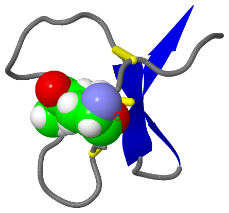
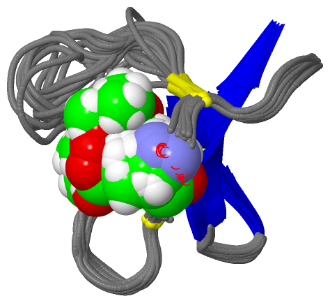
NMR Structure (1, 1)
No.NameCountTypeFull Name
1GME1Mod. Amino Acid5-O-METHYL-GLUTAMIC ACID

(-) Sites  (0, 0)

(no "Site" information available for 2KNN)

(-) SS Bonds  (3, 3)

NMR Structure
No.Residues
1A:4 -A:20
2A:8 -A:22
3A:13 -A:27

(-) Cis Peptide Bonds  (0, 0)

(no "Cis Peptide Bond" information available for 2KNN)

 Sequence-Structure Mapping

(-) SAPs(SNPs)/Variants  (0, 0)

(no "SAP(SNP)/Variant" information available for 2KNN)

(-) PROSITE Motifs  (2, 2)

NMR Structure (2, 2)
 PROSITEUniProtKBPDB
No.IDACDescriptionIDLocationCountLocation
1CYCLOTIDEPS51052 Cyclotides profile.CYO2_VIOOD1-30  1A:1-30
2CYCLOTIDE_BRACELETPS60008 Cyclotides bracelet subfamily signature.CYO2_VIOOD4-13  1A:4-13

(-) Exons   (0, 0)

(no "Exon" information available for 2KNN)

(-) Sequences/Alignments

NMR Structure
   Reformat: Number of residues per line =  ('0' or empty: single-line sequence representation)
  Number of residues per labelling interval =   
  UniProt sequence: complete  aligned part    
   Show mapping: SCOP domains CATH domains Pfam domains Secondary structure (by author)
SAPs(SNPs) PROSITE motifs Exons
(details for a mapped element are shown in a popup box when the mouse pointer rests over it)
Chain A from PDB  Type:PROTEIN  Length:30
 aligned with CYO2_VIOOD | P58434 from UniProtKB/Swiss-Prot  Length:30

    Alignment length:30
                                    10        20        30
            CYO2_VIOOD    1 GIPCGESCVWIPCISSAIGCSCKSKVCYRN 30
               SCOP domains d2knna_ A: automated matches   SCOP domains
               CATH domains ------------------------------ CATH domains
               Pfam domains Cyclotide-2knnA01 A:1-30       Pfam domains
         Sec.struct. author .eeeeee.............eee..eeee. Sec.struct. author
                 SAPs(SNPs) ------------------------------ SAPs(SNPs)
                PROSITE (1) CYCLOTIDE  PDB: A:1-30         PROSITE (1)
                PROSITE (2) ---CYCLOTIDE_----------------- PROSITE (2)
                 Transcript ------------------------------ Transcript
                  2knn A  1 GIPCGeSCVWIPCISSAIGCSCKSKVCYRN 30
                                 |  10        20        30
                                 6-GME                    

   Legend:   → Mismatch (orange background)
  - → Gap (green background, '-', border residues have a numbering label)
    → Modified Residue (blue background, lower-case, 'x' indicates undefined single-letter code, labelled with number + name)
  x → Chemical Group (purple background, 'x', labelled with number + name, e.g. ACE or NH2)
  extra numbering lines below/above indicate numbering irregularities and modified residue names etc., number ends below/above '|'

 Classification and Annotation

(-) SCOP Domains  (1, 1)

NMR Structure

(-) CATH Domains  (0, 0)

(no "CATH Domain" information available for 2KNN)

(-) Pfam Domains  (1, 1)

NMR Structure

(-) Gene Ontology  (5, 5)

NMR Structure(hide GO term definitions)
Chain A   (CYO2_VIOOD | P58434)
molecular function
    GO:0003674    molecular_function    Elemental activities, such as catalysis or binding, describing the actions of a gene product at the molecular level. A given gene product may exhibit one or more molecular functions.
biological process
    GO:0019835    cytolysis    The rupture of cell membranes and the loss of cytoplasm.
    GO:0006952    defense response    Reactions, triggered in response to the presence of a foreign body or the occurrence of an injury, which result in restriction of damage to the organism attacked or prevention/recovery from the infection caused by the attack.
    GO:0044179    hemolysis in other organism    The cytolytic destruction of red blood cells, with the release of intracellular hemoglobin, in one organism by another.
cellular component
    GO:0005575    cellular_component    The part of a cell, extracellular environment or virus in which a gene product is located. A gene product may be located in one or more parts of a cell and its location may be as specific as a particular macromolecular complex, that is, a stable, persistent association of macromolecules that function together.

 Visualization

(-) Interactive Views

NMR Structure
  Complete Structure
    Jena3D(integrated viewing of ligand, site, SAP, PROSITE, SCOP information)
    WebMol | AstexViewer[tm]@PDBe
(Java Applets, require no local installation except for Java; loading may be slow)
    STRAP
(Java WebStart application, automatic local installation, requires Java; full application with system access!)
    RasMol
(require local installation)
    Molscript (VRML)
(requires installation of a VRML viewer; select preferred view via VRML and generate a mono or stereo PDF format file)
 
  Ligands, Modified Residues, Ions
    GME  [ RasMol | Jena3D ]  +environment [ RasMol | Jena3D ]
 
  Sites
(no "Sites" information available for 2knn)
 
  Cis Peptide Bonds
(no "Cis Peptide Bonds" information available for 2knn)
 

(-) Still Images

Jmol
  protein: cartoon or spacefill or dots and stick; nucleic acid: cartoon and stick; ligands: spacefill; active site: stick
Molscript
  protein, nucleic acid: cartoon; ligands: spacefill; active site: ball and stick

 Databases and Analysis Tools

(-) Databases

Access by PDB/NDB ID
  2knn
    Family and Domain InformationProDom | SYSTERS
    General Structural InformationGlycoscienceDB | MMDB | NDB | OCA | PDB | PDBe | PDBj | PDBsum | PDBWiki | PQS | PROTEOPEDIA
    Orientation in MembranesOPM
    Protein SurfaceSURFACE
    Secondary StructureDSSP (structure derived) | HSSP (homology derived)
    Structural GenomicsGeneCensus
    Structural NeighboursCE | VAST
    Structure ClassificationCATH | Dali | SCOP
    Validation and Original DataBMRB Data View | BMRB Restraints Grid | EDS | PROCHECK | RECOORD | WHAT_CHECK
 
Access by UniProt ID/Accession number
  CYO2_VIOOD | P58434
    Comparative Protein Structure ModelsModBase
    Genomic InformationEnsembl
    Protein-protein InteractionDIP
    Sequence, Family and Domain InformationInterPro | Pfam | SMART | UniProtKB/SwissProt
 
Access by Enzyme Classificator   (EC Number)
  (no 'Enzyme Classificator' available)
    General Enzyme InformationBRENDA | EC-PDB | Enzyme | IntEnz
    PathwayKEGG | MetaCyc
 
Access by Disease Identifier   (MIM ID)
  (no 'MIM ID' available)
    Disease InformationOMIM
 
Access by GenAge ID
  (no 'GenAge ID' available)
    Age Related InformationGenAge

(-) Analysis Tools

Access by PDB/NDB ID
    Domain InformationXDom
    Interatomic Contacts of Structural UnitsCSU
    Ligand-protein ContactsLPC
    Protein CavitiescastP
    Sequence and Secondary StructurePDBCartoon
    Structure AlignmentSTRAP(Java WebStart application, automatic local installation, requires Java; full application with system access!)
    Structure and Sequence BrowserSTING
 
Access by UniProt ID/Accession number
  CYO2_VIOOD | P58434
    Protein Disorder PredictionDisEMBL | FoldIndex | GLOBPLOT (for more information see DisProt)

 Related Entries

(-) Entries Sharing at Least One Protein Chain (UniProt ID)

UniProtKB/Swiss-Prot
        CYO2_VIOOD | P584342kcg 2knm

(-) Related Entries Specified in the PDB File

2knm NMR STRUCTURE OF THE NATIVE UNMODIFIED PEPTIDE.