Show PDB file:   
         Plain Text   HTML   (compressed file size)
QuickSearch:   
by PDB,NDB,UniProt,PROSITE Code or Search Term(s)  
(-)NMR Structure - model 1
(-)NMR Structure - all models
collapse expand < >
Image NMR Structure - model 1
NMR Structure - model 1  (Jmol Viewer)
Image NMR Structure - all models
NMR Structure - all models  (Jmol Viewer)

(-) Description

Title :  SOLUTION STRUCTURE OF PROTEIN YLBL (BSU15050) FROM BACILLUS SUBTILIS, NORTHEAST STRUCTURAL GENOMICS CONSORTIUM TARGET SR713A
 
Authors :  Y. Liu, R. Belote, C. Ciccosanti, K. Hamilton, R. Nair, B. Rost, T. Acton, R. Xiao, G. Swapna, J. Everett, G. T. Montelione, J. Prestegard, Northeast Structural Genomics Consortium (Nesg)
Date :  05 Jun 09  (Deposition) - 25 Aug 09  (Release) - 25 Aug 09  (Revision)
Method :  SOLUTION NMR
Resolution :  NOT APPLICABLE
Chains :  NMR Structure  :  A  (21x)
Keywords :  Mixed Alpha-Beta Protein, Cell Membrane, Hydrolase, Membrane, Protease, Serine Protease, Transmembrane, Structural Genomics, Psi-2, Protein Structure Initiative, Northeast Structural Genomics Consortium, Nesg, Unknown Function (Keyword Search: [Gene Ontology, PubMed, Web (Google))
 
Reference :  Y. Liu, R. Belote, C. Ciccosanti, K. Hamilton, R. Nair, B. Rost, T. Acton, R. Xiao, G. Swapna, J. Everett, G. T. Montelione, J. Prestegard
Solution Structure Of Protein Ylbl (Bsu15050) From Bacillus Subtilis, Northeast Structural Genomics Consortium Target Sr713A
To Be Published
PubMed: search
(for further references see the PDB file header)

(-) Compounds

Molecule 1 - UNCHARACTERIZED PROTEIN YLBL
    ChainsA
    EngineeredYES
    Expression SystemESCHERICHIA COLI
    Expression System Taxid562
    Expression System VectorPET 21-23C
    FragmentSEQUENCE DATABASE RESIDUES 128-210
    GeneYLBL, BSU15050
    Organism ScientificBACILLUS SUBTILIS
    Organism Taxid1423

 Structural Features

(-) Chains, Units

  
NMR Structure (21x)

Summary Information (see also Sequences/Alignments below)

(-) Ligands, Modified Residues, Ions  (0, 0)

(no "Ligand,Modified Residues,Ions" information available for 2KJP)

(-) Sites  (0, 0)

(no "Site" information available for 2KJP)

(-) SS Bonds  (0, 0)

(no "SS Bond" information available for 2KJP)

(-) Cis Peptide Bonds  (0, 0)

(no "Cis Peptide Bond" information available for 2KJP)

 Sequence-Structure Mapping

(-) SAPs(SNPs)/Variants  (0, 0)

(no "SAP(SNP)/Variant" information available for 2KJP)

(-) PROSITE Motifs  (1, 1)

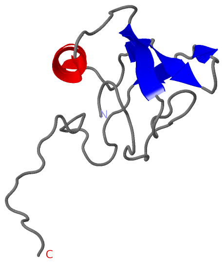
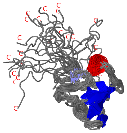
NMR Structure (1, 1)
 PROSITEUniProtKBPDB
No.IDACDescriptionIDLocationCountLocation
1LON_PROTEOLYTICPS51786 Lon proteolytic domain profile.YLBL_BACSU187-336  1A:60-84

(-) Exons   (0, 0)

(no "Exon" information available for 2KJP)

(-) Sequences/Alignments

NMR Structure
   Reformat: Number of residues per line =  ('0' or empty: single-line sequence representation)
  Number of residues per labelling interval =   
  UniProt sequence: complete  aligned part    
   Show mapping: SCOP domains CATH domains Pfam domains Secondary structure (by author)
SAPs(SNPs) PROSITE motifs Exons
(details for a mapped element are shown in a popup box when the mouse pointer rests over it)
Chain A from PDB  Type:PROTEIN  Length:91
 aligned with YLBL_BACSU | O34470 from UniProtKB/Swiss-Prot  Length:341

    Alignment length:91
                                   137       147       157       167       177       187       197       207       217 
           YLBL_BACSU   128 NGIYASSVVENMPAKGKIEVGDKIISADGKNYQSAEKLIDYISSKKAGDKVTLKIEREEKEKRVTLTLKQFPDEPDRAGIGVSLYTDRNVK 218
               SCOP domains ------------------------------------------------------------------------------------------- SCOP domains
               CATH domains ------------------------------------------------------------------------------------------- CATH domains
               Pfam domains ------------------------------------------------------------------------------------------- Pfam domains
         Sec.struct. author ......................eeeee......hhhhhhhhhh......ee.eeee....ee...ee........................ Sec.struct. author
                 SAPs(SNPs) ------------------------------------------------------------------------------------------- SAPs(SNPs)
                    PROSITE -----------------------------------------------------------LON_PROTEOLYTIC  PDB: A:60-84    PROSITE
                 Transcript ------------------------------------------------------------------------------------------- Transcript
                 2kjp A   1 NGIYASSVVENMPAKGKIEVGDKIISADGKNYQSAEKLIDYISSKKAGDKVTLKIEREEKEKRVTLTLKQFPDEPDRAGIGVSLEHHHHHH  91
                                    10        20        30        40        50        60        70        80        90 

   Legend:   → Mismatch (orange background)
  - → Gap (green background, '-', border residues have a numbering label)
    → Modified Residue (blue background, lower-case, 'x' indicates undefined single-letter code, labelled with number + name)
  x → Chemical Group (purple background, 'x', labelled with number + name, e.g. ACE or NH2)
  extra numbering lines below/above indicate numbering irregularities and modified residue names etc., number ends below/above '|'

 Classification and Annotation

(-) SCOP Domains  (0, 0)

(no "SCOP Domain" information available for 2KJP)

(-) CATH Domains  (0, 0)

(no "CATH Domain" information available for 2KJP)

(-) Pfam Domains  (0, 0)

(no "Pfam Domain" information available for 2KJP)

(-) Gene Ontology  (11, 11)

NMR Structure(hide GO term definitions)
Chain A   (YLBL_BACSU | O34470)
molecular function
    GO:0005524    ATP binding    Interacting selectively and non-covalently with ATP, adenosine 5'-triphosphate, a universally important coenzyme and enzyme regulator.
    GO:0004176    ATP-dependent peptidase activity    Catalysis of the reaction: ATP + H2O = ADP + phosphate, to drive the hydrolysis of peptide bonds.
    GO:0016787    hydrolase activity    Catalysis of the hydrolysis of various bonds, e.g. C-O, C-N, C-C, phosphoric anhydride bonds, etc. Hydrolase is the systematic name for any enzyme of EC class 3.
    GO:0008233    peptidase activity    Catalysis of the hydrolysis of a peptide bond. A peptide bond is a covalent bond formed when the carbon atom from the carboxyl group of one amino acid shares electrons with the nitrogen atom from the amino group of a second amino acid.
    GO:0004252    serine-type endopeptidase activity    Catalysis of the hydrolysis of internal, alpha-peptide bonds in a polypeptide chain by a catalytic mechanism that involves a catalytic triad consisting of a serine nucleophile that is activated by a proton relay involving an acidic residue (e.g. aspartate or glutamate) and a basic residue (usually histidine).
    GO:0008236    serine-type peptidase activity    Catalysis of the hydrolysis of peptide bonds in a polypeptide chain by a catalytic mechanism that involves a catalytic triad consisting of a serine nucleophile that is activated by a proton relay involving an acidic residue (e.g. aspartate or glutamate) and a basic residue (usually histidine).
biological process
    GO:0030163    protein catabolic process    The chemical reactions and pathways resulting in the breakdown of a protein by the destruction of the native, active configuration, with or without the hydrolysis of peptide bonds.
    GO:0006508    proteolysis    The hydrolysis of proteins into smaller polypeptides and/or amino acids by cleavage of their peptide bonds.
cellular component
    GO:0016021    integral component of membrane    The component of a membrane consisting of the gene products and protein complexes having at least some part of their peptide sequence embedded in the hydrophobic region of the membrane.
    GO:0016020    membrane    A lipid bilayer along with all the proteins and protein complexes embedded in it an attached to it.
    GO:0005886    plasma membrane    The membrane surrounding a cell that separates the cell from its external environment. It consists of a phospholipid bilayer and associated proteins.

 Visualization

(-) Interactive Views

NMR Structure
  Complete Structure
    Jena3D(integrated viewing of ligand, site, SAP, PROSITE, SCOP information)
    WebMol | AstexViewer[tm]@PDBe
(Java Applets, require no local installation except for Java; loading may be slow)
    STRAP
(Java WebStart application, automatic local installation, requires Java; full application with system access!)
    RasMol
(require local installation)
    Molscript (VRML)
(requires installation of a VRML viewer; select preferred view via VRML and generate a mono or stereo PDF format file)
 
  Ligands, Modified Residues, Ions
(no "Ligands, Modified Residues, Ions" information available for 2kjp)
 
  Sites
(no "Sites" information available for 2kjp)
 
  Cis Peptide Bonds
(no "Cis Peptide Bonds" information available for 2kjp)
 

(-) Still Images

Jmol
  protein: cartoon or spacefill or dots and stick; nucleic acid: cartoon and stick; ligands: spacefill; active site: stick
Molscript
  protein, nucleic acid: cartoon; ligands: spacefill; active site: ball and stick

 Databases and Analysis Tools

(-) Databases

Access by PDB/NDB ID
  2kjp
    Family and Domain InformationProDom | SYSTERS
    General Structural InformationGlycoscienceDB | MMDB | NDB | OCA | PDB | PDBe | PDBj | PDBsum | PDBWiki | PQS | PROTEOPEDIA
    Orientation in MembranesOPM
    Protein SurfaceSURFACE
    Secondary StructureDSSP (structure derived) | HSSP (homology derived)
    Structural GenomicsGeneCensus
    Structural NeighboursCE | VAST
    Structure ClassificationCATH | Dali | SCOP
    Validation and Original DataBMRB Data View | BMRB Restraints Grid | EDS | PROCHECK | RECOORD | WHAT_CHECK
 
Access by UniProt ID/Accession number
  YLBL_BACSU | O34470
    Comparative Protein Structure ModelsModBase
    Genomic InformationEnsembl
    Protein-protein InteractionDIP
    Sequence, Family and Domain InformationInterPro | Pfam | SMART | UniProtKB/SwissProt
 
Access by Enzyme Classificator   (EC Number)
  (no 'Enzyme Classificator' available)
    General Enzyme InformationBRENDA | EC-PDB | Enzyme | IntEnz
    PathwayKEGG | MetaCyc
 
Access by Disease Identifier   (MIM ID)
  (no 'MIM ID' available)
    Disease InformationOMIM
 
Access by GenAge ID
  (no 'GenAge ID' available)
    Age Related InformationGenAge

(-) Analysis Tools

Access by PDB/NDB ID
    Domain InformationXDom
    Interatomic Contacts of Structural UnitsCSU
    Ligand-protein ContactsLPC
    Protein CavitiescastP
    Sequence and Secondary StructurePDBCartoon
    Structure AlignmentSTRAP(Java WebStart application, automatic local installation, requires Java; full application with system access!)
    Structure and Sequence BrowserSTING
 
Access by UniProt ID/Accession number
  YLBL_BACSU | O34470
    Protein Disorder PredictionDisEMBL | FoldIndex | GLOBPLOT (for more information see DisProt)

 Related Entries

(-) Entries Sharing at Least One Protein Chain (UniProt ID)

(no "Entries Sharing at Least One Protein Chain" available for 2KJP)

(-) Related Entries Specified in the PDB File

(no "Related Entries Specified in the PDB File" available for 2KJP)