|
|
|
|
 Description
|
|
Compounds
|
||||||||||||||||||||||||||||||||||||||||||||||||||||||||||||||||||||||||||
Chains, Units
Summary Information (see also Sequences/Alignments below) |
Ligands, Modified Residues, Ions (0, 0)| (no "Ligand,Modified Residues,Ions" information available for 2KJ4) |
Sites (0, 0)| (no "Site" information available for 2KJ4) |
SS Bonds (3, 3)
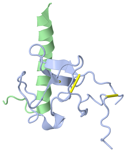
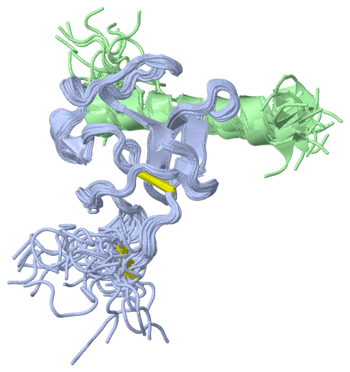
NMR Structure
|
||||||||||||||||
Cis Peptide Bonds (1, 20)
NMR Structure
|
||||||||||
SAPs(SNPs)/Variants (0, 0)| (no "SAP(SNP)/Variant" information available for 2KJ4) |
PROSITE Motifs (0, 0)| (no "PROSITE Motif" information available for 2KJ4) |
Exons (0, 0)| (no "Exon" information available for 2KJ4) |
Sequences/Alignments
NMR Structure
Chain A from PDB Type:PROTEIN Length:87
SCOP domains d2kj4a_ A: automated matches SCOP domains
CATH domains --------------------------------------------------------------------------------------- CATH domains
Pfam domains --------------------------------------------------------------------------------------- Pfam domains
SAPs(SNPs) --------------------------------------------------------------------------------------- SAPs(SNPs)
PROSITE --------------------------------------------------------------------------------------- PROSITE
Transcript --------------------------------------------------------------------------------------- Transcript
2kj4 A 1 YVEFSEECMHGSGENYDGKISKTMSGLECQAWDSQSPHAHGYIPSKFPNKNLKKNYCRNPDRDLRPWCFTTDPNKRWEYCDIPRCAA 87
10 20 30 40 50 60 70 80
Chain B from PDB Type:PROTEIN Length:32 aligned with Q6V4L8_STRPY | Q6V4L8 from UniProtKB/TrEMBL Length:388 Alignment length:32 92 102 112 Q6V4L8_STRPY 83 DDVEKLTADAELQRLKNERHEEAELERLKSER 114 SCOP domains -------------------------------- SCOP domains CATH domains -------------------------------- CATH domains Pfam domains (1) ---------------------VEK-30-2kj- Pfam domains (1) Pfam domains (2) ---------------------VEK-30-2kj- Pfam domains (2) SAPs(SNPs) -------------------------------- SAPs(SNPs) PROSITE -------------------------------- PROSITE Transcript -------------------------------- Transcript 2kj4 B 99 GSVEKLTADAELQRLKNERHEEAELERLKSEY 130 108 118 128
|
||||||||||||||||||||
SCOP Domains (1, 1)
NMR Structure
 |
CATH Domains (0, 0)| (no "CATH Domain" information available for 2KJ4) |
Pfam Domains (1, 2)
NMR Structure
|
Gene Ontology (1, 1)|
NMR Structure(hide GO term definitions) Chain B (Q6V4L8_STRPY | Q6V4L8)
|
||||||||||||
Interactive Views
|
|||||||||||||||||||||||||||||||||||||||||||||||||||||||||||||||||||||||||||||||||||||||||||||||||||||||||||||||||||||
Still Images
|
||||||||||||||||
Databases
|
||||||||||||||||||||||||||||||||||||||||||||||||||||||||||||||||||||||||||||||||||||||||||||||||||||||||||||||||||||||||||||||||||||||||||||||||||||||||||||||||
Analysis Tools
|
|||||||||||||||||||||||||||||||||||||||||||||||||||||||||||||
Entries Sharing at Least One Protein Chain (UniProt ID)
Related Entries Specified in the PDB File
|
|