|
|
|
|
 Description
|
|
Compounds
|
||||||||||||||||||||||||
Chains, Units
Summary Information (see also Sequences/Alignments below) |
Ligands, Modified Residues, Ions (0, 0)| (no "Ligand,Modified Residues,Ions" information available for 2K84) |
Sites (0, 0)| (no "Site" information available for 2K84) |
SS Bonds (0, 0)| (no "SS Bond" information available for 2K84) |
Cis Peptide Bonds (0, 0)| (no "Cis Peptide Bond" information available for 2K84) |
SAPs(SNPs)/Variants (0, 0)| (no "SAP(SNP)/Variant" information available for 2K84) |
PROSITE Motifs (0, 0)| (no "PROSITE Motif" information available for 2K84) |
Exons (0, 0)| (no "Exon" information available for 2K84) |
Sequences/Alignments
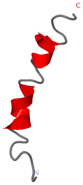
NMR StructureChain A from PDB Type:PROTEIN Length:30 aligned with GAG_EIAVY | P69732 from UniProtKB/Swiss-Prot Length:486 Alignment length:30 466 476 486 GAG_EIAVY 457 LYPDLSEIKKEYNVKEKDQVEDLNLDSLWE 486 SCOP domains ------------------------------ SCOP domains CATH domains ------------------------------ CATH domains Pfam domains ------------------------------ Pfam domains SAPs(SNPs) ------------------------------ SAPs(SNPs) PROSITE ------------------------------ PROSITE Transcript ------------------------------ Transcript 2k84 A 22 LYPDLSEIKKEYNVKEKDQVEDLNLDSLWE 51 31 41 51
|
||||||||||||||||||||
SCOP Domains (0, 0)| (no "SCOP Domain" information available for 2K84) |
CATH Domains (0, 0)| (no "CATH Domain" information available for 2K84) |
Pfam Domains (0, 0)| (no "Pfam Domain" information available for 2K84) |
Gene Ontology (12, 12)|
NMR Structure(hide GO term definitions) Chain A (GAG_EIAVY | P69732)
|
||||||||||||||||||||||||||||||||||||||||||||||||||||||||||||||||||||||||||||||||||||||||||
Interactive Views
|
||||||||||||||||||||||||||||||||||||||||||||||||||||||||||||||||||||||||||||||||||||||||||||||||||||||||||||||||||||
Still Images
|
||||||||||||||||
Databases
|
||||||||||||||||||||||||||||||||||||||||||||||||||||||||||||||||||||||||||||||||||||||||||||||||||||||||||||||||||||||||||||||||||||||||||||||||||||||||||||||||
Analysis Tools
|
|||||||||||||||||||||||||||||||||||||||||||||||||||||||||||||
Entries Sharing at Least One Protein Chain (UniProt ID)
Related Entries Specified in the PDB File
|
|