|
|
|
|
 Description
|
|
Compounds
|
||||||||||||||||||||||||||||||||||||||||||||||||||||||||
Chains, Units
Summary Information (see also Sequences/Alignments below) |
Ligands, Modified Residues, Ions (0, 0)| (no "Ligand,Modified Residues,Ions" information available for 2K1G) |
Sites (0, 0)| (no "Site" information available for 2K1G) |
SS Bonds (0, 0)| (no "SS Bond" information available for 2K1G) |
Cis Peptide Bonds (0, 0)| (no "Cis Peptide Bond" information available for 2K1G) |
SAPs(SNPs)/Variants (0, 0)| (no "SAP(SNP)/Variant" information available for 2K1G) |
PROSITE Motifs (0, 0)| (no "PROSITE Motif" information available for 2K1G) |
Exons (0, 0)| (no "Exon" information available for 2K1G) |
Sequences/Alignments
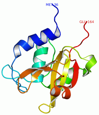
NMR StructureChain A from PDB Type:PROTEIN Length:129 aligned with MEPS_ECOLI | P0AFV4 from UniProtKB/Swiss-Prot Length:188 Alignment length:129 188 71 81 91 101 111 121 131 141 151 161 171 181 | MEPS_ECOLI 62 RNVDVKSRIMDQYADWKGVRYRLGGSTKKGIDCSGFVQRTFREQFGLELPRSTYEQQEMGKSVSRSNLRTGDLVLFRAGSTGRHVGIYIGNNQFVHASTSSGVIISSMNEPYWKKRYNEARRVLSRS-- - SCOP domains --------------------------------------------------------------------------------------------------------------------------------- SCOP domains CATH domains 2k1gA00 A:36-164 endopeptidase domain like (from Nostoc punctiforme) CATH domains Pfam domains --------------------------------------------------------------------------------------------------------------------------------- Pfam domains SAPs(SNPs) --------------------------------------------------------------------------------------------------------------------------------- SAPs(SNPs) PROSITE --------------------------------------------------------------------------------------------------------------------------------- PROSITE Transcript --------------------------------------------------------------------------------------------------------------------------------- Transcript 2k1g A 36 MNVDVKSRIMDQYADWKGVRYRLGGSTKKGIDCSGFVQRTFREQFGLELPRSTYEQQEMGKSVSRSNLRTGDLVLFRAGSTGRHVGIYIGNNQFVHASTSSGVIISSMNEPYWKKRYNEARRVLSRSLE 164 45 55 65 75 85 95 105 115 125 135 145 155
|
||||||||||||||||||||
SCOP Domains (0, 0)| (no "SCOP Domain" information available for 2K1G) |
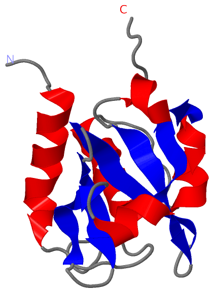
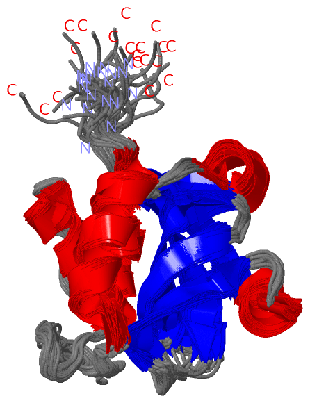
CATH Domains (1, 1)
NMR Structure
 |
Pfam Domains (0, 0)| (no "Pfam Domain" information available for 2K1G) |
Gene Ontology (12, 12)|
NMR Structure(hide GO term definitions) Chain A (MEPS_ECOLI | P0AFV4)
|
||||||||||||||||||||||||||||||||||||||||||||||||||||||||||||||||||||||||||||||||||||||||||
Interactive Views
|
||||||||||||||||||||||||||||||||||||||||||||||||||||||||||||||||||||||||||||||||||||||||||||||||||||||||||||||||||||
Still Images
|
||||||||||||||||
Databases
|
||||||||||||||||||||||||||||||||||||||||||||||||||||||||||||||||||||||||||||||||||||||||||||||||||||||||||||||||||||||||||||||||||||||||||||||||||||||||||||||||
Analysis Tools
|
|||||||||||||||||||||||||||||||||||||||||||||||||||||||||||||
Entries Sharing at Least One Protein Chain (UniProt ID)
Related Entries Specified in the PDB File
|
|