|
|
|
|
 Description
|
|
Compounds
|
||||||||||||||||
Chains, Units
Summary Information (see also Sequences/Alignments below) |
Ligands, Modified Residues, Ions (0, 0)| (no "Ligand,Modified Residues,Ions" information available for 2JR8) |
Sites (0, 0)| (no "Site" information available for 2JR8) |
SS Bonds (0, 0)| (no "SS Bond" information available for 2JR8) |
Cis Peptide Bonds (0, 0)| (no "Cis Peptide Bond" information available for 2JR8) |
SAPs(SNPs)/Variants (0, 0)| (no "SAP(SNP)/Variant" information available for 2JR8) |
PROSITE Motifs (0, 0)| (no "PROSITE Motif" information available for 2JR8) |
Exons (0, 0)| (no "Exon" information available for 2JR8) |
Sequences/Alignments
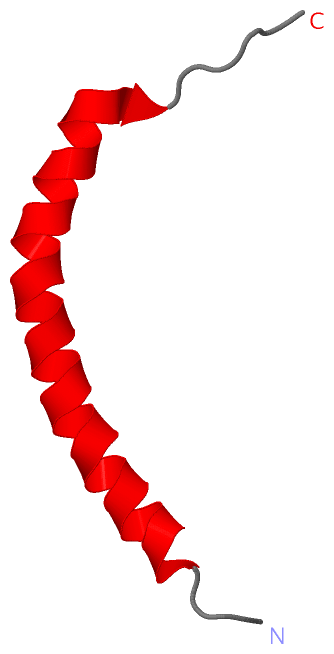
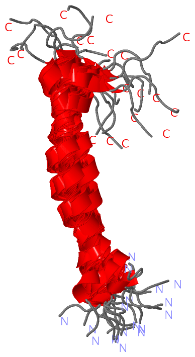
NMR StructureChain A from PDB Type:PROTEIN Length:42 aligned with Q86MA1_MANSE | Q86MA1 from UniProtKB/TrEMBL Length:67 Alignment length:42 35 45 55 65 Q86MA1_MANSE 26 GKIPVKAIKQAGKVIGKGLRAINIAGTTHDVVSFFRPKKKKH 67 SCOP domains -d2jr8a1 A:2-41 Moricin - SCOP domains CATH domains 2jr8A00 A:1-42 Single helix bin CATH domains Pfam domains Moricin-2jr8A01 A:1-41 - Pfam domains SAPs(SNPs) ------------------------------------------ SAPs(SNPs) PROSITE ------------------------------------------ PROSITE Transcript ------------------------------------------ Transcript 2jr8 A 1 GKIPVKAIKQAGKVIGKGLRAINIAGTTHDVVSFFRPKKKKH 42 10 20 30 40
|
||||||||||||||||||||
SCOP Domains (1, 1)
NMR Structure
 |
CATH Domains (1, 1)
NMR Structure
|
Pfam Domains (1, 1)
NMR Structure
|
Gene Ontology (2, 2)|
NMR Structure(hide GO term definitions) Chain A (Q86MA1_MANSE | Q86MA1)
|
||||||||||||||||||||||||
Interactive Views
|
||||||||||||||||||||||||||||||||||||||||||||||||||||||||||||||||||||||||||||||||||||||||||||||||||||||||||||||||||||
Still Images
|
||||||||||||||||
Databases
|
||||||||||||||||||||||||||||||||||||||||||||||||||||||||||||||||||||||||||||||||||||||||||||||||||||||||||||||||||||||||||||||||||||||||||||||||||||||||||||||||
Analysis Tools
|
|||||||||||||||||||||||||||||||||||||||||||||||||||||||||||||
Entries Sharing at Least One Protein Chain (UniProt ID)
Related Entries Specified in the PDB File
|
|