| molecular function |
|---|
| | GO:0005254 | | chloride channel activity | | Enables the facilitated diffusion of a chloride (by an energy-independent process) involving passage through a transmembrane aqueous pore or channel without evidence for a carrier-mediated mechanism. |
| | GO:0005216 | | ion channel activity | | Enables the facilitated diffusion of an ion (by an energy-independent process) by passage through a transmembrane aqueous pore or channel without evidence for a carrier-mediated mechanism. May be either selective (it enables passage of a specific ion only) or non-selective (it enables passage of two or more ions of same charge but different size). |
| | GO:0044325 | | ion channel binding | | Interacting selectively and non-covalently with one or more specific sites on an ion channel, a protein complex that spans a membrane and forms a water-filled channel across the phospholipid bilayer allowing selective ion transport down its electrochemical gradient. |
| | GO:0017080 | | sodium channel regulator activity | | Modulates the activity of a sodium channel. |
| biological process |
|---|
| | GO:1902476 | | chloride transmembrane transport | | The directed movement of chloride across a membrane. |
| | GO:0006821 | | chloride transport | | The directed movement of chloride into, out of or within a cell, or between cells, by means of some agent such as a transporter or pore. |
| | GO:0034220 | | ion transmembrane transport | | A process in which an ion is transported from one side of a membrane to the other by means of some agent such as a transporter or pore. |
| | GO:0006811 | | ion transport | | The directed movement of charged atoms or small charged molecules into, out of or within a cell, or between cells, by means of some agent such as a transporter or pore. |
| | GO:0006936 | | muscle contraction | | A process in which force is generated within muscle tissue, resulting in a change in muscle geometry. Force generation involves a chemo-mechanical energy conversion step that is carried out by the actin/myosin complex activity, which generates force through ATP hydrolysis. |
| | GO:1903278 | | positive regulation of sodium ion export from cell | | Any process that activates or increases the frequency, rate or extent of sodium ion export from cell. |
| | GO:1903779 | | regulation of cardiac conduction | | Any process that modulates the frequency, rate or extent of cardiac conduction. |
| | GO:0086036 | | regulation of cardiac muscle cell membrane potential | | Any process that modulates the establishment or extent of a membrane potential in a cardiac muscle cell (a cardiomyocyte). A membrane potential is the electric potential existing across any membrane arising from charges in the membrane itself and from the charges present in the media on either side of the membrane. |
| | GO:0008016 | | regulation of heart contraction | | Any process that modulates the frequency, rate or extent of heart contraction. Heart contraction is the process in which the heart decreases in volume in a characteristic way to propel blood through the body. |
| | GO:2000649 | | regulation of sodium ion transmembrane transporter activity | | Any process that modulates the frequency, rate or extent of sodium ion transmembrane transporter activity. |
| | GO:0006810 | | transport | | The directed movement of substances (such as macromolecules, small molecules, ions) or cellular components (such as complexes and organelles) into, out of or within a cell, or between cells, or within a multicellular organism by means of some agent such as a transporter, pore or motor protein. |
| cellular component |
|---|
| | GO:0034707 | | chloride channel complex | | An ion channel complex through which chloride ions pass. |
| | GO:0016021 | | integral component of membrane | | The component of a membrane consisting of the gene products and protein complexes having at least some part of their peptide sequence embedded in the hydrophobic region of the membrane. |
| | GO:0005887 | | integral component of plasma membrane | | The component of the plasma membrane consisting of the gene products and protein complexes having at least some part of their peptide sequence embedded in the hydrophobic region of the membrane. |
| | GO:0016020 | | membrane | | A lipid bilayer along with all the proteins and protein complexes embedded in it an attached to it. |
| | GO:0005886 | | plasma membrane | | The membrane surrounding a cell that separates the cell from its external environment. It consists of a phospholipid bilayer and associated proteins. |
| | GO:0042383 | | sarcolemma | | The outer membrane of a muscle cell, consisting of the plasma membrane, a covering basement membrane (about 100 nm thick and sometimes common to more than one fiber), and the associated loose network of collagen fibers. |
| | GO:0005890 | | sodium:potassium-exchanging ATPase complex | | Sodium:potassium-exchanging ATPases are tetrameric proteins, consisting of two large alpha subunits and two smaller beta subunits. The alpha subunits bear the active site and penetrate the membrane, while the beta subunits carry oligosaccharide groups and face the cell exterior. |
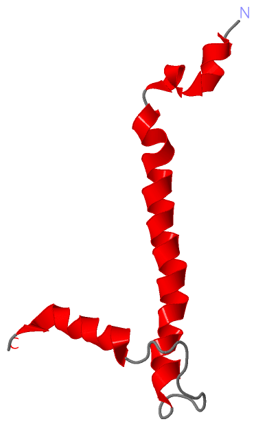
 Description
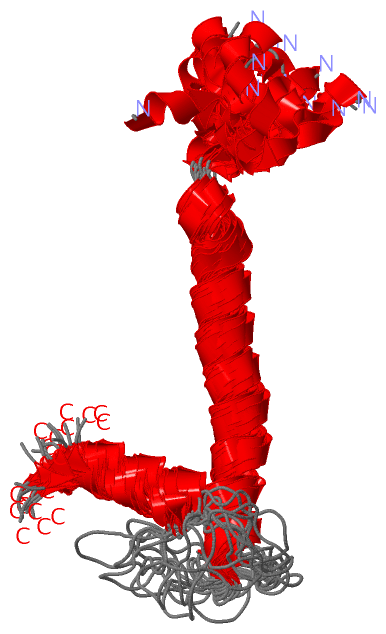