Show PDB file:   
         Plain Text   HTML   (compressed file size)
QuickSearch:   
by PDB,NDB,UniProt,PROSITE Code or Search Term(s)  
(-)NMR Structure - model 1
(-)NMR Structure - model 1, sites
(-)NMR Structure - all models
collapse expand < >
Image NMR Structure - model 1
NMR Structure - model 1  (Jmol Viewer)
Image NMR Structure - model 1, sites
NMR Structure - model 1, sites  (Jmol Viewer)
Image NMR Structure - all models
NMR Structure - all models  (Jmol Viewer)

(-) Description

Title :  NMR DERIVED SOLUTION STRUCTURE OF AN EF-HAND CALCIUM BINDING PROTEIN FROM ENTAMOEBA HISTOLYTICA
 
Authors :  S. M. Mustafi, R. B. Mutalik, K. V. Chary
Date :  11 Feb 07  (Deposition) - 12 Feb 08  (Release) - 24 Feb 09  (Revision)
Method :  SOLUTION NMR
Resolution :  NOT APPLICABLE
Chains :  NMR Structure  :  A  (20x)
Keywords :  Calcium Binding Protein, Ehcabp2, Metal Binding Protein (Keyword Search: [Gene Ontology, PubMed, Web (Google))
 
Reference :  S. M. Mustafi, R. B. Mutalik
Structure, Dynamics, And Physiological Properties Of A Calcium Binding Protein From Entamoeba Histolytica (Ehcabp2)
To Be Published
PubMed: search
(for further references see the PDB file header)

(-) Compounds

Molecule 1 - CALCIUM BINDING PROTEIN 2
    ChainsA
    EngineeredYES
    Expression SystemESCHERICHIA COLI
    Expression System Taxid562
    Organism ScientificENTAMOEBA HISTOLYTICA
    Organism Taxid5759

 Structural Features

(-) Chains, Units

  
NMR Structure (20x)

Summary Information (see also Sequences/Alignments below)

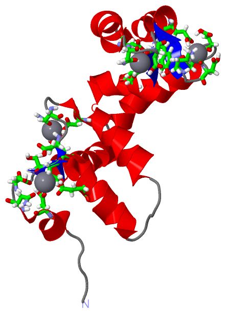
(-) Ligands, Modified Residues, Ions  (1, 4)

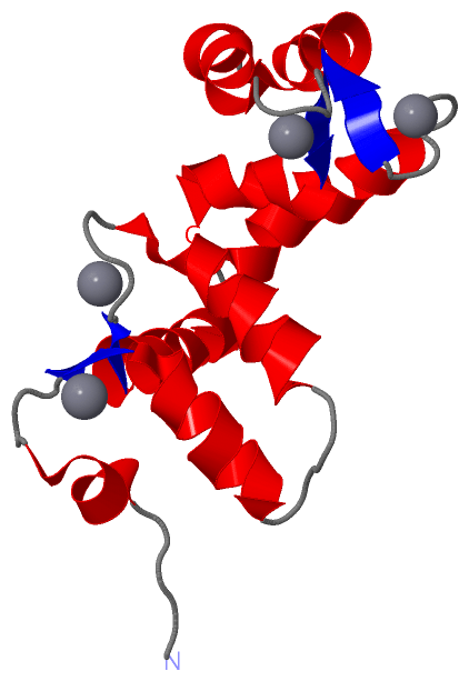
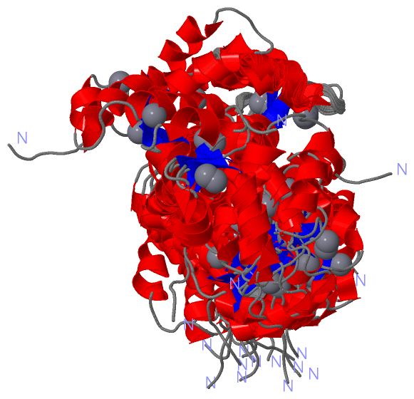
NMR Structure (1, 4)
No.NameCountTypeFull Name
1CA4Ligand/IonCALCIUM ION

(-) Sites  (4, 4)

NMR Structure (4, 4)
No.NameEvidenceResiduesDescription
1AC1SOFTWAREASP A:10 , ASN A:12 , ASP A:14 , SER A:16 , VAL A:17 , GLU A:21BINDING SITE FOR RESIDUE CA A 145
2AC2SOFTWAREASP A:46 , ASN A:50 , GLU A:52 , GLU A:57BINDING SITE FOR RESIDUE CA A 155
3AC3SOFTWAREASP A:85 , ASP A:87 , GLY A:88 , ASP A:89 , LYS A:91 , GLU A:96BINDING SITE FOR RESIDUE CA A 175
4AC4SOFTWAREASP A:117 , ASN A:119 , ASP A:121 , TYR A:123 , GLU A:128BINDING SITE FOR RESIDUE CA A 185

(-) SS Bonds  (0, 0)

(no "SS Bond" information available for 2JNX)

(-) Cis Peptide Bonds  (0, 0)

(no "Cis Peptide Bond" information available for 2JNX)

 Sequence-Structure Mapping

(-) SAPs(SNPs)/Variants  (0, 0)

(no "SAP(SNP)/Variant" information available for 2JNX)

(-) PROSITE Motifs  (0, 0)

(no "PROSITE Motif" information available for 2JNX)

(-) Exons   (0, 0)

(no "Exon" information available for 2JNX)

(-) Sequences/Alignments

NMR Structure
   Reformat: Number of residues per line =  ('0' or empty: single-line sequence representation)
  Number of residues per labelling interval =   
  UniProt sequence: complete  aligned part    
   Show mapping: SCOP domains CATH domains Pfam domains Secondary structure (by author)
SAPs(SNPs) PROSITE motifs Exons
(details for a mapped element are shown in a popup box when the mouse pointer rests over it)
Chain A from PDB  Type:PROTEIN  Length:134
 aligned with Q6R3G0_ENTHI | Q6R3G0 from UniProtKB/TrEMBL  Length:134

    Alignment length:134
                                    10        20        30        40        50        60        70        80        90       100       110       120       130    
         Q6R3G0_ENTHI     1 MAEALFKQLDANGDGSVSYEEVKAFVSSKRPIKNEQLLQLIFKAIDIDGNGEIDLAEFTKFAAAVKEQDLSDEKVGLKILYKLMDADGDGKLTKEEVTTFFKKFGYEKVVDQIMKADANGDGYITLEEFLAFNL 134
               SCOP domains d2jnxa_ A: automated matches                                                                                                           SCOP domains
               CATH domains -------------------------------------------------------------------------------------------------------------------------------------- CATH domains
           Pfam domains (1) -----------------------------------------------------------------EF_hand_5-2jnxA01 A:66-132                                         -- Pfam domains (1)
           Pfam domains (2) -----------------------------------------------------------------EF_hand_5-2jnxA02 A:66-132                                         -- Pfam domains (2)
           Pfam domains (3) -------------------------------------------------EF_hand_6-2jnxA03 A:50-104                             ------------------------------ Pfam domains (3)
           Pfam domains (4) -------------------------------------------------EF_hand_6-2jnxA04 A:50-104                             ------------------------------ Pfam domains (4)
         Sec.struct. author ....hhhhhhh....eehhhhhhhhhhh.....hhhhhhhhhhhhhh.....eehhhhhhhhhhhhh..hhhhhhhhhhhhhhh......eeehhhhhhhhhhhh..hhhhhhhhhh.....eeehhhhhhhh. Sec.struct. author
                 SAPs(SNPs) -------------------------------------------------------------------------------------------------------------------------------------- SAPs(SNPs)
                    PROSITE -------------------------------------------------------------------------------------------------------------------------------------- PROSITE
                 Transcript -------------------------------------------------------------------------------------------------------------------------------------- Transcript
                 2jnx A   1 MAEALFKQLDANGDGSVSYEEVKAFVSSKRPIKNEQLLQLIFKAIDIDGNGEIDLAEFTKFAAAVKEQDLSDEKVGLKILYKLMDADGDGKLTKEEVTTFFKKFGYEKVVDQIMKADANGDGYITLEEFLAFNL 134
                                    10        20        30        40        50        60        70        80        90       100       110       120       130    

   Legend:   → Mismatch (orange background)
  - → Gap (green background, '-', border residues have a numbering label)
    → Modified Residue (blue background, lower-case, 'x' indicates undefined single-letter code, labelled with number + name)
  x → Chemical Group (purple background, 'x', labelled with number + name, e.g. ACE or NH2)
  extra numbering lines below/above indicate numbering irregularities and modified residue names etc., number ends below/above '|'

 Classification and Annotation

(-) SCOP Domains  (1, 1)

NMR Structure

(-) CATH Domains  (0, 0)

(no "CATH Domain" information available for 2JNX)

(-) Pfam Domains  (2, 4)

NMR Structure
(-)
Clan: EF_hand (270)

(-) Gene Ontology  (2, 2)

NMR Structure(hide GO term definitions)
Chain A   (Q6R3G0_ENTHI | Q6R3G0)
molecular function
    GO:0005509    calcium ion binding    Interacting selectively and non-covalently with calcium ions (Ca2+).
    GO:0046872    metal ion binding    Interacting selectively and non-covalently with any metal ion.

 Visualization

(-) Interactive Views

NMR Structure
  Complete Structure
    Jena3D(integrated viewing of ligand, site, SAP, PROSITE, SCOP information)
    WebMol | AstexViewer[tm]@PDBe
(Java Applets, require no local installation except for Java; loading may be slow)
    STRAP
(Java WebStart application, automatic local installation, requires Java; full application with system access!)
    RasMol
(require local installation)
    Molscript (VRML)
(requires installation of a VRML viewer; select preferred view via VRML and generate a mono or stereo PDF format file)
 
  Ligands, Modified Residues, Ions
    CA  [ RasMol | Jena3D ]  +environment [ RasMol | Jena3D ]
 
  Sites
    AC1  [ RasMol ]  +environment [ RasMol ]
    AC2  [ RasMol ]  +environment [ RasMol ]
    AC3  [ RasMol ]  +environment [ RasMol ]
    AC4  [ RasMol ]  +environment [ RasMol ]
 
  Cis Peptide Bonds
(no "Cis Peptide Bonds" information available for 2jnx)
 

(-) Still Images

Jmol
  protein: cartoon or spacefill or dots and stick; nucleic acid: cartoon and stick; ligands: spacefill; active site: stick
Molscript
  protein, nucleic acid: cartoon; ligands: spacefill; active site: ball and stick

 Databases and Analysis Tools

(-) Databases

Access by PDB/NDB ID
  2jnx
    Family and Domain InformationProDom | SYSTERS
    General Structural InformationGlycoscienceDB | MMDB | NDB | OCA | PDB | PDBe | PDBj | PDBsum | PDBWiki | PQS | PROTEOPEDIA
    Orientation in MembranesOPM
    Protein SurfaceSURFACE
    Secondary StructureDSSP (structure derived) | HSSP (homology derived)
    Structural GenomicsGeneCensus
    Structural NeighboursCE | VAST
    Structure ClassificationCATH | Dali | SCOP
    Validation and Original DataBMRB Data View | BMRB Restraints Grid | EDS | PROCHECK | RECOORD | WHAT_CHECK
 
Access by UniProt ID/Accession number
  Q6R3G0_ENTHI | Q6R3G0
    Comparative Protein Structure ModelsModBase
    Genomic InformationEnsembl
    Protein-protein InteractionDIP
    Sequence, Family and Domain InformationInterPro | Pfam | SMART | UniProtKB/TrEMBL
 
Access by Enzyme Classificator   (EC Number)
  (no 'Enzyme Classificator' available)
    General Enzyme InformationBRENDA | EC-PDB | Enzyme | IntEnz
    PathwayKEGG | MetaCyc
 
Access by Disease Identifier   (MIM ID)
  (no 'MIM ID' available)
    Disease InformationOMIM
 
Access by GenAge ID
  (no 'GenAge ID' available)
    Age Related InformationGenAge

(-) Analysis Tools

Access by PDB/NDB ID
    Domain InformationXDom
    Interatomic Contacts of Structural UnitsCSU
    Ligand-protein ContactsLPC
    Protein CavitiescastP
    Sequence and Secondary StructurePDBCartoon
    Structure AlignmentSTRAP(Java WebStart application, automatic local installation, requires Java; full application with system access!)
    Structure and Sequence BrowserSTING
 
Access by UniProt ID/Accession number
  Q6R3G0_ENTHI | Q6R3G0
    Protein Disorder PredictionDisEMBL | FoldIndex | GLOBPLOT (for more information see DisProt)

 Related Entries

(-) Entries Sharing at Least One Protein Chain (UniProt ID)

(no "Entries Sharing at Least One Protein Chain" available for 2JNX)

(-) Related Entries Specified in the PDB File

2jn12jn2
6433