|
|
|
|
 Description
|
|
Compounds
|
||||||||||||||||||||||||||||||||||||||||||||
Chains, Units
Summary Information (see also Sequences/Alignments below) |
Ligands, Modified Residues, Ions (0, 0)| (no "Ligand,Modified Residues,Ions" information available for 2JNF) |
Sites (0, 0)| (no "Site" information available for 2JNF) |
SS Bonds (0, 0)| (no "SS Bond" information available for 2JNF) |
Cis Peptide Bonds (0, 0)| (no "Cis Peptide Bond" information available for 2JNF) |
SAPs(SNPs)/Variants (0, 0)| (no "SAP(SNP)/Variant" information available for 2JNF) |
PROSITE Motifs (0, 0)| (no "PROSITE Motif" information available for 2JNF) |
Exons (0, 0)| (no "Exon" information available for 2JNF) |
Sequences/Alignments
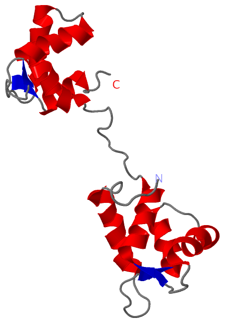
NMR StructureChain A from PDB Type:PROTEIN Length:158 aligned with Q868D4_9HEMI | Q868D4 from UniProtKB/TrEMBL Length:158 Alignment length:158 10 20 30 40 50 60 70 80 90 100 110 120 130 140 150 Q868D4_9HEMI 1 MGDVSKLSSNQVKLLETAFRDFETPEGSGRVSTDQIGIILEVLGIQQTKSTIRQLIDEFDPFGNGDIDFDSFKIIGARFLGEEVNPEQMQQELREAFRLYDKEGNGYISTDVMREILAELDETLSSEDLDAMIDEIDADGSGTVDFEEFMGVMTGGDE 158 SCOP domains d2jnfa_ A: automated matches SCOP domains CATH domains -------------------------------------------------------------------------------------------------------------------------------------------------------------- CATH domains Pfam domains (1) -------------------------------------EF_hand_6-2jnfA02 A:38-79 ------------EF_hand_5-2jnfA01 A:92-153 ----- Pfam domains (1) Pfam domains (2) -------------------------------------------------------------------------------------------------------------------------------efhand-2jnfA03 A:128-156 -- Pfam domains (2) SAPs(SNPs) -------------------------------------------------------------------------------------------------------------------------------------------------------------- SAPs(SNPs) PROSITE -------------------------------------------------------------------------------------------------------------------------------------------------------------- PROSITE Transcript -------------------------------------------------------------------------------------------------------------------------------------------------------------- Transcript 2jnf A 1 MGDVSKLSSNQVKLLETAFRDFETPEGSGRVSTDQIGIILEVLGIQQTKSTIRQLIDEFDPFGNGDIDFDSFKIIGARFLGEEVNPEQMQQELREAFRLYDKEGNGYISTDVMREILAELDETLSSEDLDAMIDEIDADGSGTVDFEEFMGVMTGGDE 158 10 20 30 40 50 60 70 80 90 100 110 120 130 140 150
|
||||||||||||||||||||
SCOP Domains (1, 1)
NMR Structure
 |
CATH Domains (0, 0)| (no "CATH Domain" information available for 2JNF) |
Pfam Domains (3, 3)
NMR Structure
|
Gene Ontology (1, 1)|
NMR Structure(hide GO term definitions) Chain A (Q868D4_9HEMI | Q868D4)
|
||||||||||||
Interactive Views
|
||||||||||||||||||||||||||||||||||||||||||||||||||||||||||||||||||||||||||||||||||||||||||||||||||||||||||||||||||||
Still Images
|
||||||||||||||||
Databases
|
||||||||||||||||||||||||||||||||||||||||||||||||||||||||||||||||||||||||||||||||||||||||||||||||||||||||||||||||||||||||||||||||||||||||||||||||||||||||||||||||
Analysis Tools
|
|||||||||||||||||||||||||||||||||||||||||||||||||||||||||||||
Entries Sharing at Least One Protein Chain (UniProt ID)
Related Entries Specified in the PDB File
|
|