Show PDB file:   
         Plain Text   HTML   (compressed file size)
QuickSearch:   
by PDB,NDB,UniProt,PROSITE Code or Search Term(s)  
(-)Asymmetric Unit
(-)Asym. Unit - sites
(-)Biological Unit 1
(-)Biol. Unit 1 - sites
collapse expand < >
Image Asymmetric Unit
Asymmetric Unit  (Jmol Viewer)
Image Asym. Unit - sites
Asym. Unit - sites  (Jmol Viewer)
Image Biological Unit 1
Biological Unit 1  (Jmol Viewer)
Image Biol. Unit 1 - sites
Biol. Unit 1 - sites  (Jmol Viewer)

(-) Description

Title :  CRYSTAL STRUCTURE OF RECOMBINANT DIOCLEA GUIANENSIS LECTIN S131H COMPLEXED WITH 5-BROMO-4-CHLORO-3-INDOLYL-A-D-MANNOSE
 
Authors :  C. S. Nagano, L. Sanz, B. S. Cavada, J. J. Calvete
Date :  15 Jan 07  (Deposition) - 30 Oct 07  (Release) - 13 Apr 11  (Revision)
Method :  X-RAY DIFFRACTION
Resolution :  1.65
Chains :  Asym. Unit :  A
Biol. Unit 1:  A  (4x)
Keywords :  Carbohydrate Binding Protein, Cona-Like, Metal-Binding, Legume Lectin, Recombinant Lectin, Sugar-Binding Protein (Keyword Search: [Gene Ontology, PubMed, Web (Google))
 
Reference :  C. S. Nagano, J. J. Calvete, D. Barettino, A. Perez, B. S. Cavada, L. San
Insights Into The Structural Basis Of The Ph- Dependent Dimer-Tetramer Equilibrium Through Crystallographic Analysis Of Recombinant Diocleinae Lectins.
Biochem. J. V. 409 417 2008
PubMed-ID: 17937659  |  Reference-DOI: 10.1042/BJ20070942

(-) Compounds

Molecule 1 - LECTIN ALPHA CHAIN
    ChainsA
    EngineeredYES
    Expression SystemESCHERICHIA COLI
    Expression System PlasmidRDGUIAS131H/PET32A
    Expression System StrainBL21(DE3)
    Expression System Taxid469008
    Expression System VectorPET32A
    Expression System Vector TypePLASMID
    MutationYES
    Organism ScientificDIOCLEA GUIANENSIS
    Organism Taxid99571
    SynonymRDGUIA

 Structural Features

(-) Chains, Units

  1
Asymmetric Unit A
Biological Unit 1 (4x)A

Summary Information (see also Sequences/Alignments below)
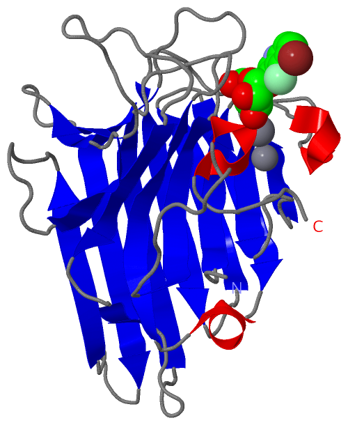
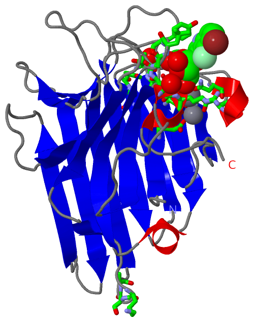
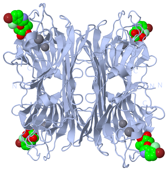
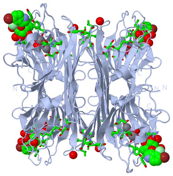
(-) Ligands, Modified Residues, Ions  (3, 3)

Asymmetric Unit (3, 3)
No.NameCountTypeFull Name
1CA1Ligand/IonCALCIUM ION
2MN1Ligand/IonMANGANESE (II) ION
3XMM1Ligand/Ion(2R,3S,4S,5S,6R)-2-(5-BROMO-4-CHLORO-1H-INDOL-3-YLOXY)-TETRAHYDRO-6-(HYDROXYMETHYL)-2H-PYRAN-3,4,5-TRIOL
Biological Unit 1 (1, 4)
No.NameCountTypeFull Name
1CA-1Ligand/IonCALCIUM ION
2MN-1Ligand/IonMANGANESE (II) ION
3XMM4Ligand/Ion(2R,3S,4S,5S,6R)-2-(5-BROMO-4-CHLORO-1H-INDOL-3-YLOXY)-TETRAHYDRO-6-(HYDROXYMETHYL)-2H-PYRAN-3,4,5-TRIOL

(-) Sites  (3, 3)

Asymmetric Unit (3, 3)
No.NameEvidenceResiduesDescription
1AC1SOFTWAREGLU A:10 , ASP A:12 , ASP A:21 , HIS A:26 , HOH A:2252 , HOH A:2253BINDING SITE FOR RESIDUE MN A1240
2AC2SOFTWAREASP A:12 , TYR A:14 , ASN A:16 , ASP A:21 , HOH A:2221 , HOH A:2240BINDING SITE FOR RESIDUE CA A1241
3AC3SOFTWARETYR A:14 , ASN A:16 , SER A:68 , TYR A:69 , THR A:72 , SER A:73 , GLY A:100 , LEU A:101 , TYR A:102 , LYS A:186 , SER A:187 , ALA A:188 , ALA A:209 , ASP A:210 , GLY A:229 , ARG A:230 , HOH A:2094 , HOH A:2254 , HOH A:2255BINDING SITE FOR RESIDUE XMM A1242

(-) SS Bonds  (0, 0)

(no "SS Bond" information available for 2JE7)

(-) Cis Peptide Bonds  (1, 1)

Asymmetric Unit
No.Residues
1Ala A:209 -Asp A:210

 Sequence-Structure Mapping

(-) SAPs(SNPs)/Variants  (0, 0)

(no "SAP(SNP)/Variant" information available for 2JE7)

(-) PROSITE Motifs  (2, 2)

Asymmetric Unit (2, 2)
 PROSITEUniProtKBPDB
No.IDACDescriptionIDLocationCountLocation
1LECTIN_LEGUME_BETAPS00307 Legume lectins beta-chain signature.LECA_DIOGU5-11  1A:7-13
2LECTIN_LEGUME_ALPHAPS00308 Legume lectins alpha-chain signature.LECA_DIOGU85-94  1A:87-96
Biological Unit 1 (2, 8)
 PROSITEUniProtKBPDB
No.IDACDescriptionIDLocationCountLocation
1LECTIN_LEGUME_BETAPS00307 Legume lectins beta-chain signature.LECA_DIOGU5-11  4A:7-13
2LECTIN_LEGUME_ALPHAPS00308 Legume lectins alpha-chain signature.LECA_DIOGU85-94  4A:87-96

(-) Exons   (0, 0)

(no "Exon" information available for 2JE7)

(-) Sequences/Alignments

Asymmetric Unit
   Reformat: Number of residues per line =  ('0' or empty: single-line sequence representation)
  Number of residues per labelling interval =   
  UniProt sequence: complete  aligned part    
   Show mapping: SCOP domains CATH domains Pfam domains Secondary structure (by author)
SAPs(SNPs) PROSITE motifs Exons
(details for a mapped element are shown in a popup box when the mouse pointer rests over it)
Chain A from PDB  Type:PROTEIN  Length:237
 aligned with LECA_DIOGU | P81637 from UniProtKB/Swiss-Prot  Length:237

    Alignment length:237
                                    10        20        30        40        50        60        70        80        90       100       110       120       130       140       150       160       170       180       190       200       210       220       230       
           LECA_DIOGU     1 ADTIVAVELDSYPNTDIGDPSYPHIGIDIKSIRSKSTARWNMQTGKVGTAHISYNSVAKRLSAVVSYTGSSSTTVSYDVDLNNVLPEWVRVGLSATTGLYKETNTILSWSFTSKLKTNSIADANSLHFSFNQFSQNPKDLILQSDATTDSDGNLELTKVSSSGDPQGSSVGRALFYAPVHIWEKSAVVAGFDATFTFLIKSPDRDPADGITFFIANTDTSIPSGSGGRLLGLFPDAN 237
               SCOP domains d2je7a_ A: automated matches                                                                                                                                                                                                                  SCOP domains
               CATH domains 2je7A00 A:3-239  [code=2.60.120.200, no name defined]                                                                                                                                                                                         CATH domains
           Pfam domains (1) ---------------------------------------------------------------------------------------------------------------------------Lectin_legB-2je7A01 A:126-239                                                                                      Pfam domains (1)
           Pfam domains (2) ---------------------------------------------------------------------------------------------------------------------------Lectin_legB-2je7A02 A:126-239                                                                                      Pfam domains (2)
         Sec.struct. author ...eeeeeee...hhhhh.....eeeeee......eeee.......eeeeeeeee....eeeeeeee...eeeeeeee.hhhhhh..eeeeeeeee........eeeeeeeeeeee.......eeeeeee.........eeeee..ee.....ee..............eeeeee...ee.......eeeeeeeeeee..........eeeeeeee.........hhhhh....... Sec.struct. author
                 SAPs(SNPs) --------------------------------------------------------------------------------------------------------------------------------------------------------------------------------------------------------------------------------------------- SAPs(SNPs)
                    PROSITE ----LECTIN_-------------------------------------------------------------------------LECTIN_LEG----------------------------------------------------------------------------------------------------------------------------------------------- PROSITE
                 Transcript --------------------------------------------------------------------------------------------------------------------------------------------------------------------------------------------------------------------------------------------- Transcript
                 2je7 A   3 ADTIVAVELDSYPNTDIGDPSYPHIGIDIKSIRSKSTARWNMQTGKVGTAHISYNSVAKRLSAVVSYSGTSSTTVSYDVDLNNVLPEWVRVGLSATTGLYKETNTILSWSFTSKLKTNSIADANSLHFSFHQFSQNPKDLILQSDATTDSDGNLQLTRVSSDGSPQGSSVGRALFYAPVHIWEKSAVVASFDATFTFLIKSPDRDPADGITFFIANTDTSIPSGSGGRLLGLFPDAN 239
                                    12        22        32        42        52        62        72        82        92       102       112       122       132       142       152       162       172       182       192       202       212       222       232       

   Legend:   → Mismatch (orange background)
  - → Gap (green background, '-', border residues have a numbering label)
    → Modified Residue (blue background, lower-case, 'x' indicates undefined single-letter code, labelled with number + name)
  x → Chemical Group (purple background, 'x', labelled with number + name, e.g. ACE or NH2)
  extra numbering lines below/above indicate numbering irregularities and modified residue names etc., number ends below/above '|'

 Classification and Annotation

(-) SCOP Domains  (1, 1)

Asymmetric Unit

(-) CATH Domains  (1, 1)

Asymmetric Unit
(-)
Class: Mainly Beta (13760)

(-) Pfam Domains  (1, 2)

Asymmetric Unit

(-) Gene Ontology  (5, 5)

Asymmetric Unit(hide GO term definitions)
Chain A   (LECA_DIOGU | P81637)
molecular function
    GO:0030246    carbohydrate binding    Interacting selectively and non-covalently with any carbohydrate, which includes monosaccharides, oligosaccharides and polysaccharides as well as substances derived from monosaccharides by reduction of the carbonyl group (alditols), by oxidation of one or more hydroxy groups to afford the corresponding aldehydes, ketones, or carboxylic acids, or by replacement of one or more hydroxy group(s) by a hydrogen atom. Cyclitols are generally not regarded as carbohydrates.
    GO:0005537    mannose binding    Interacting selectively and non-covalently with mannose, a monosaccharide hexose, stereoisomeric with glucose, that occurs naturally only in polymerized forms called mannans.
    GO:0046872    metal ion binding    Interacting selectively and non-covalently with any metal ion.
biological process
    GO:0008150    biological_process    Any process specifically pertinent to the functioning of integrated living units: cells, tissues, organs, and organisms. A process is a collection of molecular events with a defined beginning and end.
cellular component
    GO:0005575    cellular_component    The part of a cell, extracellular environment or virus in which a gene product is located. A gene product may be located in one or more parts of a cell and its location may be as specific as a particular macromolecular complex, that is, a stable, persistent association of macromolecules that function together.

 Visualization

(-) Interactive Views

Asymmetric Unit
  Complete Structure
    Jena3D(integrated viewing of ligand, site, SAP, PROSITE, SCOP information)
    WebMol | AstexViewer[tm]@PDBe
(Java Applets, require no local installation except for Java; loading may be slow)
    STRAP
(Java WebStart application, automatic local installation, requires Java; full application with system access!)
    RasMol
(require local installation)
    Molscript (VRML)
(requires installation of a VRML viewer; select preferred view via VRML and generate a mono or stereo PDF format file)
 
  Ligands, Modified Residues, Ions
    CA  [ RasMol | Jena3D ]  +environment [ RasMol | Jena3D ]
    MN  [ RasMol | Jena3D ]  +environment [ RasMol | Jena3D ]
    XMM  [ RasMol | Jena3D ]  +environment [ RasMol | Jena3D ]
 
  Sites
    AC1  [ RasMol ]  +environment [ RasMol ]
    AC2  [ RasMol ]  +environment [ RasMol ]
    AC3  [ RasMol ]  +environment [ RasMol ]
 
  Cis Peptide Bonds
    Ala A:209 - Asp A:210   [ RasMol ]  
 
Biological Unit
  Complete Structure
    Biological Unit 1  [ Jena3D ]

(-) Still Images

Jmol
  protein: cartoon or spacefill or dots and stick; nucleic acid: cartoon and stick; ligands: spacefill; active site: stick
Molscript
  protein, nucleic acid: cartoon; ligands: spacefill; active site: ball and stick

 Databases and Analysis Tools

(-) Databases

Access by PDB/NDB ID
  2je7
    Family and Domain InformationProDom | SYSTERS
    General Structural InformationGlycoscienceDB | MMDB | NDB | OCA | PDB | PDBe | PDBj | PDBsum | PDBWiki | PQS | PROTEOPEDIA
    Orientation in MembranesOPM
    Protein SurfaceSURFACE
    Secondary StructureDSSP (structure derived) | HSSP (homology derived)
    Structural GenomicsGeneCensus
    Structural NeighboursCE | VAST
    Structure ClassificationCATH | Dali | SCOP
    Validation and Original DataBMRB Data View | BMRB Restraints Grid | EDS | PROCHECK | RECOORD | WHAT_CHECK
 
Access by UniProt ID/Accession number
  LECA_DIOGU | P81637
    Comparative Protein Structure ModelsModBase
    Genomic InformationEnsembl
    Protein-protein InteractionDIP
    Sequence, Family and Domain InformationInterPro | Pfam | SMART | UniProtKB/SwissProt
 
Access by Enzyme Classificator   (EC Number)
  (no 'Enzyme Classificator' available)
    General Enzyme InformationBRENDA | EC-PDB | Enzyme | IntEnz
    PathwayKEGG | MetaCyc
 
Access by Disease Identifier   (MIM ID)
  (no 'MIM ID' available)
    Disease InformationOMIM
 
Access by GenAge ID
  (no 'GenAge ID' available)
    Age Related InformationGenAge

(-) Analysis Tools

Access by PDB/NDB ID
    Domain InformationXDom
    Interatomic Contacts of Structural UnitsCSU
    Ligand-protein ContactsLPC
    Protein CavitiescastP
    Sequence and Secondary StructurePDBCartoon
    Structure AlignmentSTRAP(Java WebStart application, automatic local installation, requires Java; full application with system access!)
    Structure and Sequence BrowserSTING
 
Access by UniProt ID/Accession number
  LECA_DIOGU | P81637
    Protein Disorder PredictionDisEMBL | FoldIndex | GLOBPLOT (for more information see DisProt)

 Related Entries

(-) Entries Sharing at Least One Protein Chain (UniProt ID)

UniProtKB/Swiss-Prot
        LECA_DIOGU | P816371h9p 1h9w 2jdz

(-) Related Entries Specified in the PDB File

1h9p CRYSTAL STRUCTURE OF DIOCLEA GUIANENSIS SEED LECTIN
1h9w NATIVE DIOCLEA GUIANENSIS SEED LECTIN
2jdz CRYSTAL STRUCTURE OF RECOMBINANT DIOCLEA GUIANENSIS LECTIN COMPLEXED WITH 5-BROMO-4-CHLORO-3-INDOLYL-A-D-MANNOSE
2je9 CRYSTAL STRUCTURE OF RECOMBINANT DIOCLEA GRANDIFLORA LECTIN COMPLEXED WITH 5-BROMO-4-CHLORO-3-INDOLYL-A- D-MANNOSE
2jec CRYSTAL STRUCTURE OF RECOMBINANT DIOCLEA GRANDIFLORA LECTIN MUTANT E123A-H131N-K132Q COMPLEXED WITH 5-BROMO -4-CHLORO-3-INDOLYL-A-D-MANNOSE