|
|
|
|
 Description
|
|
Compounds
|
||||||||||||||||||||||||||||||||||||||||||||
Chains, Units
Summary Information (see also Sequences/Alignments below) |
Ligands, Modified Residues, Ions (0, 0)| (no "Ligand,Modified Residues,Ions" information available for 2JDG) |
Sites (0, 0)| (no "Site" information available for 2JDG) |
SS Bonds (0, 0)| (no "SS Bond" information available for 2JDG) |
Cis Peptide Bonds (0, 0)| (no "Cis Peptide Bond" information available for 2JDG) |
SAPs(SNPs)/Variants (3, 3)
Asymmetric/Biological Unit (3, 3)
|
|||||||||||||||||||||||||||||||||||||||||||||||||||||||||||||||||||||||||||||||
PROSITE Motifs (1, 4)
Asymmetric/Biological Unit (1, 4)
|
||||||||||||||||||||||||
Exons (3, 3)
Asymmetric/Biological Unit (3, 3)
|
||||||||||||||||||||||||||||||||||||||||||||||||||||||||||||
Sequences/Alignments
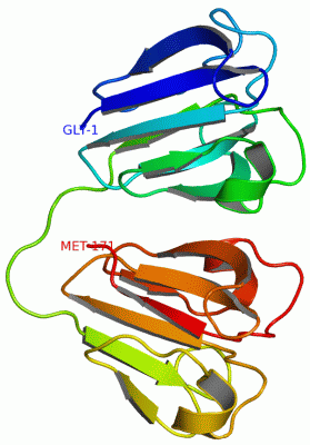
Asymmetric/Biological UnitChain A from PDB Type:PROTEIN Length:172 aligned with CRGB_HUMAN | P07316 from UniProtKB/Swiss-Prot Length:175 Alignment length:172 11 21 31 41 51 61 71 81 91 101 111 121 131 141 151 161 171 CRGB_HUMAN 2 GKITFYEDRAFQGRSYECTTDCPNLQPYFSRCNSIRVESGCWMIYERPNYQGHQYFLRRGEYPDYQQWMGLSDSIRSCCLIPPHSGAYRMKIYDRDELRGQMSELTDDCISVQDRFHLTEIHSLNVLEGSWILYEMPNYRGRQYLLRPGEYRRFLDWGAPNAKVGSLRRVMD 173 SCOP domains d2jdga1 A:1-86 automated matches -------------------------------------------------------------------------------------- SCOP domains CATH domains 2jdgA01 A:1-83 Crystallins 2jdgA02 A:84-172 Crystallins CATH domains Pfam domains (1) ----------------------------------------------------------------------------------------Crystall-2jdgA01 A:89-170 -- Pfam domains (1) Pfam domains (2) ----------------------------------------------------------------------------------------Crystall-2jdgA02 A:89-170 -- Pfam domains (2) SAPs(SNPs) -----------------------------------------------------------------------I----------------T--------------------L-------------------------------------------------------------- SAPs(SNPs) PROSITE CRYSTALLIN_BETA_GAMMA PDB: A:1-39 CRYSTALLIN_BETA_GAMMA PDB: A:40-82 -----CRYSTALLIN_BETA_GAMMA PDB: A:88-128 CRYSTALLIN_BETA_GAMMA PDB: A:129-171 - PROSITE Transcript 1 1.Exon 1.2 PDB: A:3-83 UniProt: 4-84 Exon 1.3 PDB: A:84-172 UniProt: 85-175 [INCOMPLETE] Transcript 1 2jdg A 1 GSIIFLEDRAFQGRIYGCTTDCPNLQPYFSRCNSIVVQSGCWMIYERPNYQGHQYFLRRGEYPDYQQWMGLSDSIRSCCLIPPHSGAYRMKIYDRDELRGQMSELTDDCLSVQDRFHLTEIHSLNVLEGSWILYEMPNYRGRQYLLRPGEYRRFLDWGAPNAKVGSLRRVMD 172 10 20 30 40 50 60 70 80 90 100 110 120 130 140 150 160 170
|
||||||||||||||||||||
SCOP Domains (1, 1)
Asymmetric/Biological Unit

|
CATH Domains (1, 2)
Asymmetric/Biological Unit
|
Pfam Domains (1, 2)
Asymmetric/Biological Unit
|
Gene Ontology (9, 9)|
Asymmetric/Biological Unit(hide GO term definitions) Chain A (CRGB_HUMAN | P07316)
|
||||||||||||||||||||||||||||||||||||||||||||||||||||||||||||||||||||||||
Interactive Views
|
||||||||||||||||||||||||||||||||||||||||||||||||||||||||||||||||||||||||||||||||||||||||||||||||||||||||||||||||||||
Still Images
|
||||||||||||||||
Databases
|
||||||||||||||||||||||||||||||||||||||||||||||||||||||||||||||||||||||||||||||||||||||||||||||||||||||||||||||||||||||||||||||||||||||||||||||||||||||||||||||||
Analysis Tools
|
|||||||||||||||||||||||||||||||||||||||||||||||||||||||||||||
Entries Sharing at Least One Protein Chain (UniProt ID)
Related Entries Specified in the PDB File
|
|