|
|
|
|
 Description
|
|
Compounds
|
||||||||||||||||||||||||||||||||||||||||||||||||
Chains, Units
Summary Information (see also Sequences/Alignments below) |
Ligands, Modified Residues, Ions (0, 0)| (no "Ligand,Modified Residues,Ions" information available for 2I88) |
Sites (0, 0)| (no "Site" information available for 2I88) |
SS Bonds (0, 0)| (no "SS Bond" information available for 2I88) |
Cis Peptide Bonds (0, 0)| (no "Cis Peptide Bond" information available for 2I88) |
SAPs(SNPs)/Variants (0, 0)| (no "SAP(SNP)/Variant" information available for 2I88) |
PROSITE Motifs (1, 1)
Asymmetric/Biological Unit (1, 1)
|
||||||||||||||||||||||||
Exons (0, 0)| (no "Exon" information available for 2I88) |
Sequences/Alignments
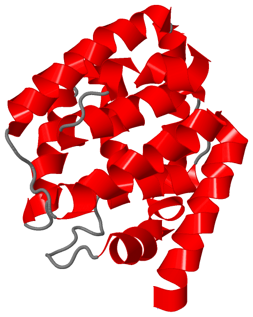
Asymmetric/Biological UnitChain A from PDB Type:PROTEIN Length:178 aligned with CEA1_ECOLX | P02978 from UniProtKB/Swiss-Prot Length:522 Alignment length:178 354 364 374 384 394 404 414 424 434 444 454 464 474 484 494 504 514 CEA1_ECOLX 345 IKDAVDATVSFYQTLTEKYGEKYSKMAQELADKSKGKKIGNVNEALAAFEKYKDVLNKKFSKADRDAIFNALASVKYDDWAKHLDQFAKYLKITGHVSFGYDVVSDILKIKDTGDWKPLFLTLEKKAADAGVSYVVALLFSLLAGTTLGIWGIAIVTGILCSYIDKNKLNTINEVLGI 522 SCOP domains ---------------------------------------------------------------------------------------------------------------------------------------------------------------------------------- SCOP domains CATH domains ---------------------------------------------------------------------------------------------------------------------------------------------------------------------------------- CATH domains Pfam domains ---------------------------------------------------------------------------------------------------------------------------------------------------------------------------------- Pfam domains SAPs(SNPs) ---------------------------------------------------------------------------------------------------------------------------------------------------------------------------------- SAPs(SNPs) PROSITE ----------------------------------------------------------------------------------------------------------------CHANNEL_COLI------------------------------------------------------ PROSITE Transcript ---------------------------------------------------------------------------------------------------------------------------------------------------------------------------------- Transcript 2i88 A 345 IKDAVDATVSFYQTLTEKYGEKYSKMAQELADKSKGKKIGNVNEALAAFEKYKDVLNKKFSKADRDAIFNALASVKYDDWAKHLDQFAKYLKITGHVSFGYDVVSDILKIKDTGDWKPLFLTLEKKAADAGVSYVVALLFSLLAGTTLGIWGIAIVTGILCSYIDKNKLNTINEVLGI 522 354 364 374 384 394 404 414 424 434 444 454 464 474 484 494 504 514
|
||||||||||||||||||||
SCOP Domains (0, 0)| (no "SCOP Domain" information available for 2I88) |
CATH Domains (0, 0)| (no "CATH Domain" information available for 2I88) |
Pfam Domains (0, 0)| (no "Pfam Domain" information available for 2I88) |
Gene Ontology (6, 6)|
Asymmetric/Biological Unit(hide GO term definitions) Chain A (CEA1_ECOLX | P02978)
|
||||||||||||||||||||||||||||||||||||||||||||||||
Interactive Views
|
||||||||||||||||||||||||||||||||||||||||||||||||||||||||||||||||||||||||||||||||||||||||||||||||||||||||||||||||||||
Still Images
|
||||||||||||||||
Databases
|
||||||||||||||||||||||||||||||||||||||||||||||||||||||||||||||||||||||||||||||||||||||||||||||||||||||||||||||||||||||||||||||||||||||||||||||||||||||||||||||||
Analysis Tools
|
|||||||||||||||||||||||||||||||||||||||||||||||||||||||||||||
Entries Sharing at Least One Protein Chain (UniProt ID)
Related Entries Specified in the PDB File
|
|