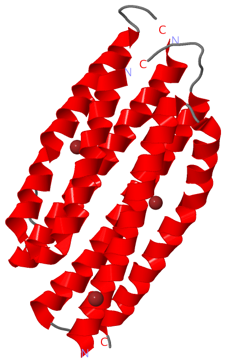
Show PDB file:   
         Plain Text   HTML   (compressed file size)
QuickSearch:   
by PDB,NDB,UniProt,PROSITE Code or Search Term(s)  
(-)Biol.Unit 1 - manually
(-)Asym.Unit - manually
(-)Asymmetric Unit
(-)Asym. Unit - sites
(-)Biological Unit 1
(-)Biol. Unit 1 - sites
(-)Biological Unit 2
collapse expand < >
Image Biol.Unit 1 - manually
Biol.Unit 1 - manually  (Jmol Viewer)
Image Asym.Unit - manually
Asym.Unit - manually  (Jmol Viewer)
Image Asymmetric Unit
Asymmetric Unit  (Jmol Viewer)
Image Asym. Unit - sites
Asym. Unit - sites  (Jmol Viewer)
Image Biological Unit 1
Biological Unit 1  (Jmol Viewer)
Image Biol. Unit 1 - sites
Biol. Unit 1 - sites  (Jmol Viewer)
Image Biological Unit 2
Biological Unit 2  (Jmol Viewer)

(-) Description

Title :  CRYSTAL STRUCTURE OF THE PHOSPHATE TRANSPORT SYSTEM REGULATORY PROTEIN PHOU FROM STREPTOCOCCUS PNEUMONIAE
 
Authors :  R. Zhang, H. Li, J. Abdullah, A. Joachimiak, Midwest Center For Stru Genomics (Mcsg)
Date :  10 Aug 06  (Deposition) - 19 Sep 06  (Release) - 13 Jul 11  (Revision)
Method :  X-RAY DIFFRACTION
Resolution :  2.40
Chains :  Asym. Unit :  A
Biol. Unit 1:  A  (2x)
Biol. Unit 2:  A  (1x)
Keywords :  Phou, Zinc-Binding Protein, Structural Genomics, Psi-2, Protein Structure Initiative, Midwest Center For Structural Genomics, Mcsg, Unknown Function (Keyword Search: [Gene Ontology, PubMed, Web (Google))
 
Reference :  R. Zhang, H. Li, J. Abdullah, A. Joachimiak
Crystal Structure Of The Phosphate Transport System Regulatory Protein Phou From Streptococcus Pneumoniae
To Be Published 2006
PubMed: search

(-) Compounds

Molecule 1 - PHOSPHATE TRANSPORT SYSTEM PROTEIN PHOU
    ChainsA
    EngineeredYES
    Expression SystemESCHERICHIA COLI BL21
    Expression System PlasmidPDM68
    Expression System StrainBL21
    Expression System Taxid511693
    Expression System Vector TypePLASMID
    GenePHOU
    Organism ScientificSTREPTOCOCCUS PNEUMONIAE
    Organism Taxid170187
    StrainTIGR4

 Structural Features

(-) Chains, Units

  1
Asymmetric Unit A
Biological Unit 1 (2x)A
Biological Unit 2 (1x)A

Summary Information (see also Sequences/Alignments below)

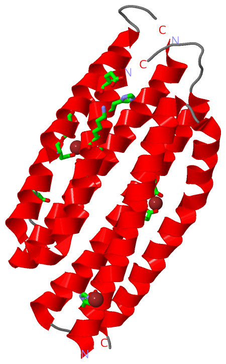
(-) Ligands, Modified Residues, Ions  (1, 3)

Asymmetric Unit (1, 3)
No.NameCountTypeFull Name
1ZN3Ligand/IonZINC ION
Biological Unit 1 (0, 0)
No.NameCountTypeFull Name
1ZN-1Ligand/IonZINC ION
Biological Unit 2 (0, 0)
No.NameCountTypeFull Name
1ZN-1Ligand/IonZINC ION

(-) Sites  (3, 3)

Asymmetric Unit (3, 3)
No.NameEvidenceResiduesDescription
1AC1SOFTWAREASP A:152 , ASP A:156 , GLU A:191 , ASP A:195BINDING SITE FOR RESIDUE ZN A 217
2AC2SOFTWAREHIS A:10 , GLU A:117 , HIS A:120 , LYS A:124BINDING SITE FOR RESIDUE ZN A 218
3AC3SOFTWAREGLU A:91 , ASP A:95 , ASP A:214BINDING SITE FOR RESIDUE ZN A 219

(-) SS Bonds  (0, 0)

(no "SS Bond" information available for 2I0M)

(-) Cis Peptide Bonds  (2, 2)

Asymmetric Unit
No.Residues
1Glu A:109 -Asn A:110
2Gln A:111 -Leu A:112

 Sequence-Structure Mapping

(-) SAPs(SNPs)/Variants  (0, 0)

(no "SAP(SNP)/Variant" information available for 2I0M)

(-) PROSITE Motifs  (0, 0)

(no "PROSITE Motif" information available for 2I0M)

(-) Exons   (0, 0)

(no "Exon" information available for 2I0M)

(-) Sequences/Alignments

Asymmetric Unit
   Reformat: Number of residues per line =  ('0' or empty: single-line sequence representation)
  Number of residues per labelling interval =   
  UniProt sequence: complete  aligned part    
   Show mapping: SCOP domains CATH domains Pfam domains Secondary structure (by author)
SAPs(SNPs) PROSITE motifs Exons
(details for a mapped element are shown in a popup box when the mouse pointer rests over it)
Chain A from PDB  Type:PROTEIN  Length:207
 aligned with PHOU_STRPN | P0A3Y7 from UniProtKB/Swiss-Prot  Length:216

    Alignment length:212
                                    13        23        33        43        53        63        73        83        93       103       113       123       133       143       153       163       173       183       193       203       213  
           PHOU_STRPN     4 QFDLELHELEQSFLGLGQLVLETASKALLALASKDKEMAELIINKDHAINQGQSAIELTCARLLALQQPQVSDLRFVISIMSSCSDLERMGDHMAGIAKAVLQLKENQLAPDEEQLHQMGKLSLSMLADLLVAFPLHQASKAISIAQKDEQIDQYYYALSKEIIGLMKDQETSIPNGTQYLYIIGHLERFADYIANICERLVYLETGELVDL 215
               SCOP domains d2i0ma_ A: automated matches                                                                                                                                                                                         SCOP domains
               CATH domains 2i0mA01 A:4-111 Phosphate transport system protein phou homolog 2;   domain 2                               2i  0mA02 A:112-215 Phosphate transport system protein phou hom olog 2; domain 2                         CATH domains
               Pfam domains -------------------------------------------------------------------------------------------------------------------------------------------------------------------------------------------------------------------- Pfam domains
         Sec.struct. author hhhhhhhhhhhhhhhhhhhhhhhhhhhhhhhhh.hhhhhhhhhhhhhhhhhhhhhhhhhhhhhh..--.hhhhhhhhhhhhhhhhhhhhhhhhhhhhhhhhhh.......--hhhhhhhhhhhhhhhhhhhhhhhhhhhhhhhhhhhhhhhhhhhhhhhhhhhhhh.....-hhhhhhhhhhhhhhhhhhhhhhhhhhhhhhhhhhh..... Sec.struct. author
                 SAPs(SNPs) -------------------------------------------------------------------------------------------------------------------------------------------------------------------------------------------------------------------- SAPs(SNPs)
                    PROSITE -------------------------------------------------------------------------------------------------------------------------------------------------------------------------------------------------------------------- PROSITE
                 Transcript -------------------------------------------------------------------------------------------------------------------------------------------------------------------------------------------------------------------- Transcript
                 2i0m A   4 QFDLELHELEQSFLGLGQLVLETASKALLALASKDKEMAELIINKDHAINQGQSAIELTCARLLAL--PQVSDLRFVISIMSSCSDLERMGDHMAGIAKAVLQLKENQLA--EEQLHQMGKLSLSMLADLLVAFPLHQASKAISIAQKDEQIDQYYYALSKEIIGLMKDQE-SIPNGTQYLYIIGHLERFADYIANICERLVYLETGELVDL 215
                                    13        23        33        43        53        63     |  73        83        93       103       113  |    123       133       143       153       163       173| |    183       193       203       213  
                                                                                            69 72                                      113  |                                                       174 |                                       
                                                                                                                                          116                                                         176                                       

   Legend:   → Mismatch (orange background)
  - → Gap (green background, '-', border residues have a numbering label)
    → Modified Residue (blue background, lower-case, 'x' indicates undefined single-letter code, labelled with number + name)
  x → Chemical Group (purple background, 'x', labelled with number + name, e.g. ACE or NH2)
  extra numbering lines below/above indicate numbering irregularities and modified residue names etc., number ends below/above '|'

 Classification and Annotation

(-) SCOP Domains  (1, 1)

Asymmetric Unit

(-) CATH Domains  (1, 2)

Asymmetric Unit

(-) Pfam Domains  (0, 0)

(no "Pfam Domain" information available for 2I0M)

(-) Gene Ontology  (7, 7)

Asymmetric Unit(hide GO term definitions)
Chain A   (PHOU_STRPN | P0A3Y7)
molecular function
    GO:0042803    protein homodimerization activity    Interacting selectively and non-covalently with an identical protein to form a homodimer.
biological process
    GO:0030643    cellular phosphate ion homeostasis    Any process involved in the maintenance of an internal steady state of phosphate ions at the level of a cell.
    GO:0045936    negative regulation of phosphate metabolic process    Any process that stops, prevents, or reduces the frequency, rate or extent of the chemical reactions and pathways involving phosphates.
    GO:2000186    negative regulation of phosphate transmembrane transport    Any process that stops, prevents, or reduces the frequency, rate or extent of phosphate transmembrane transport.
    GO:0006817    phosphate ion transport    The directed movement of phosphate into, out of or within a cell, or between cells, by means of some agent such as a transporter or pore.
    GO:0006810    transport    The directed movement of substances (such as macromolecules, small molecules, ions) or cellular components (such as complexes and organelles) into, out of or within a cell, or between cells, or within a multicellular organism by means of some agent such as a transporter, pore or motor protein.
cellular component
    GO:0005737    cytoplasm    All of the contents of a cell excluding the plasma membrane and nucleus, but including other subcellular structures.

 Visualization

(-) Interactive Views

Asymmetric Unit
  Complete Structure
    Jena3D(integrated viewing of ligand, site, SAP, PROSITE, SCOP information)
    WebMol | AstexViewer[tm]@PDBe
(Java Applets, require no local installation except for Java; loading may be slow)
    STRAP
(Java WebStart application, automatic local installation, requires Java; full application with system access!)
    RasMol
(require local installation)
    Molscript (VRML)
(requires installation of a VRML viewer; select preferred view via VRML and generate a mono or stereo PDF format file)
 
  Ligands, Modified Residues, Ions
    ZN  [ RasMol | Jena3D ]  +environment [ RasMol | Jena3D ]
 
  Sites
    AC1  [ RasMol ]  +environment [ RasMol ]
    AC2  [ RasMol ]  +environment [ RasMol ]
    AC3  [ RasMol ]  +environment [ RasMol ]
 
  Cis Peptide Bonds
    Gln A:111 - Leu A:112   [ RasMol ]  
    Glu A:109 - Asn A:110   [ RasMol ]  
 
Biological Units
  Complete Structure
    Biological Unit 1  [ Jena3D ]
    Biological Unit 2  [ Jena3D ]

(-) Still Images

Jmol
  protein: cartoon or spacefill or dots and stick; nucleic acid: cartoon and stick; ligands: spacefill; active site: stick
Molscript
  protein, nucleic acid: cartoon; ligands: spacefill; active site: ball and stick

 Databases and Analysis Tools

(-) Databases

Access by PDB/NDB ID
  2i0m
    Family and Domain InformationProDom | SYSTERS
    General Structural InformationGlycoscienceDB | MMDB | NDB | OCA | PDB | PDBe | PDBj | PDBsum | PDBWiki | PQS | PROTEOPEDIA
    Orientation in MembranesOPM
    Protein SurfaceSURFACE
    Secondary StructureDSSP (structure derived) | HSSP (homology derived)
    Structural GenomicsGeneCensus
    Structural NeighboursCE | VAST
    Structure ClassificationCATH | Dali | SCOP
    Validation and Original DataBMRB Data View | BMRB Restraints Grid | EDS | PROCHECK | RECOORD | WHAT_CHECK
 
Access by UniProt ID/Accession number
  PHOU_STRPN | P0A3Y7
    Comparative Protein Structure ModelsModBase
    Genomic InformationEnsembl
    Protein-protein InteractionDIP
    Sequence, Family and Domain InformationInterPro | Pfam | SMART | UniProtKB/SwissProt
 
Access by Enzyme Classificator   (EC Number)
  (no 'Enzyme Classificator' available)
    General Enzyme InformationBRENDA | EC-PDB | Enzyme | IntEnz
    PathwayKEGG | MetaCyc
 
Access by Disease Identifier   (MIM ID)
  (no 'MIM ID' available)
    Disease InformationOMIM
 
Access by GenAge ID
  (no 'GenAge ID' available)
    Age Related InformationGenAge

(-) Analysis Tools

Access by PDB/NDB ID
    Domain InformationXDom
    Interatomic Contacts of Structural UnitsCSU
    Ligand-protein ContactsLPC
    Protein CavitiescastP
    Sequence and Secondary StructurePDBCartoon
    Structure AlignmentSTRAP(Java WebStart application, automatic local installation, requires Java; full application with system access!)
    Structure and Sequence BrowserSTING
 
Access by UniProt ID/Accession number
  PHOU_STRPN | P0A3Y7
    Protein Disorder PredictionDisEMBL | FoldIndex | GLOBPLOT (for more information see DisProt)

 Related Entries

(-) Entries Sharing at Least One Protein Chain (UniProt ID)

(no "Entries Sharing at Least One Protein Chain" available for 2I0M)

(-) Related Entries Specified in the PDB File

1t8b 1T8B IS THE CRYSTAL STRUCTURE OF REFOLDED PHOU-LIKE PROTEIN (GI 2983430) FROM AQUIFEX AEOLICUS DETERMINED AT 3.23 ANGSTROM RESOLUTION